
Page 195 - Essential Haematology
P. 195
Chapter 13 Acute myeloid leukaemia / 181
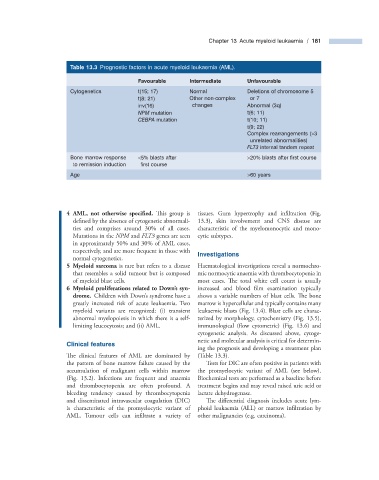
Table 13.3 Prognostic factors in acute myeloid leukaemia ( AML ).
Favourable Intermediate Unfavourable
Cytogenetics t(15; 17) Normal Deletions of chromosome 5
t(8; 21) Other non - complex or 7
inv(16) changes Abnormal (3q)
NPM mutation t(6; 11)
CEBPA mutation t(10; 11)
t(9; 22)
Complex rearrangements ( > 3
unrelated abnormalities)
FLT3 internal tandem repeat
Bone marrow response < 5% blasts after > 20% blasts after fi rst course
to remission induction fi rst course
Age > 60 years
4 AML, not otherwise specifi ed. This group is tissues. Gum hypertrophy and infi ltration (Fig.
defined by the absence of cytogenetic abnormali- 13.3 ), skin involvement and CNS disease are
ties and comprises around 30% of all cases. characteristic of the myelomonocytic and mono-
Mutations in the NPM and FLT3 genes are seen cytic subtypes.
in approximately 50% and 30% of AML cases,
respectively, and are more frequent in those with Investigations
normal cytogenetics.
5 Myeloid sarcoma is rare but refers to a disease Haematological investigations reveal a normochro-
that resembles a solid tumour but is composed mic normocytic anaemia with thrombocytopenia in
of myeloid blast cells. most cases. The total white cell count is usually
6 Myeloid proliferations related to Down ’ s syn- increased and blood film examination typically
’
drome. Children with Down s syndrome have a shows a variable numbers of blast cells. Th e bone
greatly increased risk of acute leukaemia. Two marrow is hypercellular and typically contains many
myeloid variants are recognized: (i) transient leukaemic blasts (Fig. 13.4 ). Blast cells are charac-
abnormal myelopoiesis in which there is a self - terized by morphology, cytochemistry (Fig. 13.5 ),
limiting leucocytosis; and (ii) AML. immunological (flow cytometric) (Fig. 13.6 ) and
cytogenetic analysis. As discussed above, cytoge-
netic and molecular analysis is critical for determin-
Clinical f eatures
ing the prognosis and developing a treatment plan
The clinical features of AML are dominated by (Table 13.3 ).
the pattern of bone marrow failure caused by the Tests for DIC are often positive in patients with
accumulation of malignant cells within marrow the promyelocytic variant of AML (see below).
(Fig. 13.2 ). Infections are frequent and anaemia Biochemical tests are performed as a baseline before
and thrombocytopenia are often profound. A treatment begins and may reveal raised uric acid or
bleeding tendency caused by thrombocytopenia lactate dehydrogenase.
and disseminated intravascular coagulation (DIC) Th e differential diagnosis includes acute lym-
is characteristic of the promyelocytic variant of phoid leukaemia (ALL) or marrow infi ltration by
AML. Tumour cells can infiltrate a variety of other malignancies (e.g. carcinoma).