Page 232 - Essential Haematology
P. 232

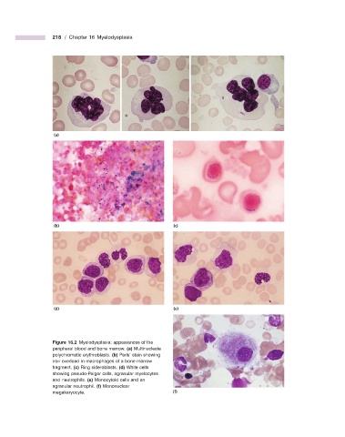
218  /  Chapter 16  Myelodysplasia


                    (a)


                    (b)                                       (c)


                    (d)                                       (e)


                              Figure 16.2   Myelodysplasia: appearances of the
                    peripheral blood and bone marrow.  (a)  Multinucleate
                    polychromatic erythroblasts.  (b)  Perls ’  stain showing
                    iron overload in macrophages of a bone marrow
                    fragment.  (c)  Ring sideroblasts.  (d)  White cells
                    showing pseudo - Pelger cells, agranular myelocytes
                    and neutrophils.  (e)  Monocytoid cells and an
                    agranular neutrophil.  (f)  Mononuclear
                    megakaryocyte.                            (f)
   227   228   229   230   231   232   233   234   235   236   237