Page 361 - Essential Haematology
P. 361

Chapter 26  Coagulation disorders  /  347


                                        X

                                                       3'           tel          3'  mRNA3'
                                                       F8B                  FVIII
                                                FVIII
                                    cen                F8A                          F8B
                                                       5'
                                                                                    F8A
                                                                                    F8A

                                                                                 5'  mRNA5'
                                    q28                F8A                  FVIII
                                                       F8A                       F8A
                                        tel         tel                        tel


                                Figure 26.2   The mechanism of the fl ip - tip inversion leading to disruption of the factor VIII gene.  (Left)  The

                      orientation of the factor VIII gene is shown with the three copies of gene A (F8A) in this region (one within an
                      intron 22 and two near the telomere).  (Middle)  During spermatogenesis at meiosis, the single X pairs with the Y
                      chromosome in the homologous regions. The X chromosome is longer than the Y and there is nothing to pair
                      with most of the long arm of X. The chromosome undergoes homologous recombination between the A genes.
                        (Right)  The fi nal result is that the factor VIII gene is disrupted. cen, centromeric end; tel, telomere; the arrows
                      indicate the direction of transcription from the A gene.


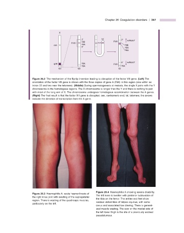
                                                                          Figure 26.4   Haemophilia A showing severe disability.
                                Figure 26.3   Haemophilia A: acute haemarthrosis of


                                                                The left knee is swollen with posterior subluxation of
                      the right knee joint with swelling of the suprapatellar
                                                                the tibia on the femur. The ankles and feet show
                      region. There is wasting of the quadriceps muscles,
                                                                residual deformities of talipes equinus, with some
                      particularly on the left.
                                                                cavus and associated toe clawing. There is general-
                                                                ized muscle wasting. The scar on the medial side of
                                                                the left lower thigh is the site of a previously excised
                                                                pseudotumour.
   356   357   358   359   360   361   362   363   364   365   366