
Page 362 - Essential Haematology
P. 362
348 / Chapter 26 Coagulation disorders
(a)
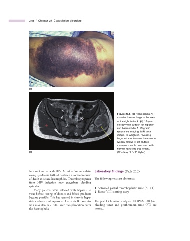
Figure 26.5 (a) Haemophilia A:
massive haemorrhage in the area
of the right buttock. (b) 15 - year -
old boy with sudden left hip pain
and haemophilia A. Magnetic
resonance imaging (MRI) axial
image, T2 weighted, revealing
large left spontaneous haematoma
(yellow arrow) in left gluteus
maximus muscle compared with
normal right side (red cross).
(b) (Courtesy of Dr P. Wylie.)
became infected with HIV. Acquired immune defi - Laboratory fi ndings (Table 26.2 )
ciency syndrome (AIDS) has been a common cause
of death in severe haemophilia. Th rombocytopenia Th e following tests are abnormal:
from HIV infection may exacerbate bleeding
episodes. 1 Activated partial thromboplastin time (APTT).
Many patients were infected with hepatitis C
virus before testing of donors and blood products 2 Factor VIII clotting assay.
became possible. This has resulted in chronic hepa-
titis, cirrhosis and hepatoma. Hepatitis B transmis- The platelet function analysis - 100 (PFA - 100) (and
sion may also be a risk. Liver transplantation cures bleeding time) and prothrombin time (PT) are
the haemophilia. normal.