
Page 373 - Essential Haematology
P. 373
Chapter 26 Coagulation disorders / 359
Coagulation d efi ciency c aused by replacement, usually as human factor VIII, recom-
a ntibodies binant VIIa or activated prothrombin complex
concentrate (FEIBA).
Circulating antibodies to coagulation factors are Another protein known as the lupus anticoagu-
occasionally seen with an incidence of approxi- lant interferes with lipoprotein - dependent stages of
mately 1 per million per year rising markedly with coagulation and is usually detected by prolongation
age. Alloantibodies to factor VIII occur in 5 – 10% of the APTT test (Table 26.6 ). This inhibitor is
of haemophiliacs. Factor VIII autoantibodies may detected in 10% of patients with systemic lupus
also result in a bleeding syndrome. Th ese immu- erythematosus (SLE) and in patients with other
noglobulin G (IgG) antibodies occur rarely post - autoimmune diseases who frequently have antibod-
partum, in certain immunological disorders (e.g. ies to other lipid - containing antigens (e.g. cardio-
rheumatoid arthritis), in cancer and in old age. lipin). The antibody is not associated with a bleeding
Treatment usually consists of a combination of tendency but there is an increased risk of arterial or
immunosuppression and treatment with factor venous thrombosis and, as with other causes of
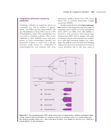
α-angle
MA A60
2 mm
20 mm
60 min
r time k time
Coagulation Fibrinolysis
Time
(a) Normal TEG trace
(b) Fibrinolysis
(c) Hypercoagulable
(d) Haemophilia
(e) Thrombocytopenia
Figure 26.11 Thromboelastography (TEG): (a – e) normal trace and appearances in different pathological states.
α - angle, speed of solid clot formation; A 60 , measure of clot lysis or retraction at 60 min; k, clot formation time;
MA, absolute strength of fi brin clot; r, rate of initial fi brin formation. (From Mallett S.V. and Cox D.J.A. (1992) Br J
Anaesth 69 ,307 – 13, with permission.)