Page 419 - Essential Haematology
P. 419
Chapter 29  Blood transfusion  /  405


                      detecting antibody or complement on the red cell             From the  p atient
                      surface where sensitization has occurred  in vivo . Th e
                                                                     1      The ABO and Rh blood group is determined.

                      AHG reagent is added to washed red cells and
                                                                   2      Serum is screened for important antibodies by an
                      agglutination indicates a positive test. A positive test
                                                                  indirect antiglobulin test on a large panel of anti-
                      occurs in haemolytic disease of the newborn,
                                                                  genically typed group O red cells.
                      autoimmune haemolytic anaemia and haemolytic
                      transfusion reactions.                        If a red cell alloantibody is discovered in the
                          The indirect antiglobulin test is used to detect   recipient, donor blood is selected lacking the rele-

                      antibodies that have coated the red cells  in vitro . It   vant antigen. The most likely are Rh D, C, c, E, e


                      is a two - stage procedure: the first step involves the   and K.
                      incubation of test red cells with serum; in the
                      second step, the red cells are washed and the AHG
                      reagent is added. Agglutination implies that the       From the  d onor
                      original serum contained antibody which has coated
                                                                  An appropriate ABO and Rh unit is selected. Donor
                      the red cells  in vitro . This test is used as part of the


                                                                (blood) testing is described on p. 399 .
                                                        ’
                      routine antibody screening of the recipient  s serum
                      prior to transfusion and for detecting blood group
                      antibodies in a pregnant woman.               The  c ross - m atch

                           Most of the above methods were originally


                      developed for tube techniques. These were replaced    The techniques that may be used are described in
                      by 96 - well microplates but most laboratories now   Table  29.8 .
                      use gel - based technology (Fig.  29.6 ).

                                                                    Electronic  c ross - m atch

                          Cross - m atching and
                                                                  In this, a patient has group and antibody screen
                        p re - t ransfusion  t ests

                                                                performed as two separate occasions. If both are
                       A number of steps are taken to ensure that patients   negative and no blood has been transfused between
                      receive compatible blood at the time of   the test, ABO and Rh compatible blood is issued
                      transfusion.                              directly without wet testing.


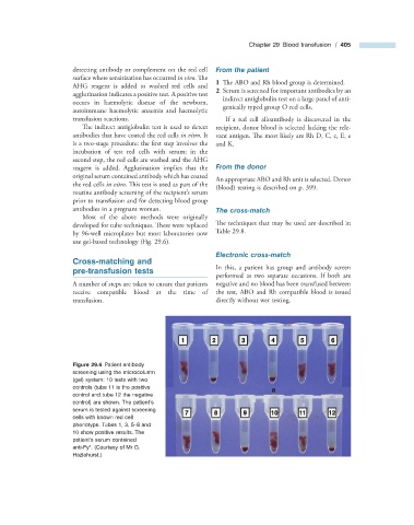
                                Figure 29.6   Patient antibody

                      screening using the microcolumn
                      (gel) system: 10 tests with two
                      controls (tube 11 is the positive
                      control and tube 12 the negative
                      control) are shown. The patient ’ s
                      serum is tested against screening
                      cells with known red cell
                      phenotype. Tubes 1, 3, 5 – 8 and
                      10 show positive results. The
                      patient ’ s serum contained
                           a
                      anti - Fy  . (Courtesy of Mr G.
                      Hazlehurst.)

