
Page 415 - Essential Haematology
P. 415
Chapter 29 Blood transfusion / 401
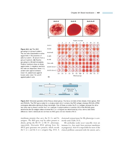
Anti-A Anti-B Anti-A+B
(a)
Patient number
1 2 3 4 5 6 7 8 9 10 11 12
Anti-A
Figure 29.3 (a) The ABO Anti-B
grouping in a group A patient.
Anti-A+B
The red cells suspended in saline
agglutinate in the presence of A cells
anti - A or anti - A + B (serum from a
group O patient). (b) Routine B cells
grouping in a 96 - well microplate.
Own cells
Positive reactions show as sharp
agglutinates; in negative reactions Anti-D clone 1
the cells are dispersed. Rows 1 – 3,
patient cells against antisera; Anti-D clone 2
rows 4 – 6, patient sera against
O+ O+ O– A+ B+ O+ AB+ B– O– AB– A+ A–
known cells; rows 7 – 8, anti - D
Blood group
against patient cells. (b)
D CcEe
Gene
Alternative
splicing
D E or e C or c
mRNA +
Figure 29.4 Molecular genetics of the Rhesus blood group. The locus consists of two closely linked genes, RhD
and RhCcEe . The RhD gene codes for a single protein which contains the RhD antigen whereas RhCcEe mRNA
undergoes alternative splicing to three transcripts. One of these encodes the E or e antigen whereas the other
two (only one is shown) contain the C or c epitope. A polymorphism at position 226 of the RhCcEe gene
determines the Ee antigen status whereas the C or c antigens are determined by a four amino acid allelic
difference. Some individuals do not have an RhD gene and are therefore Rh D – .
membrane proteins that carry the D, Cc and Ee shortened nomenclature for Rh phenotype is com-
antigens. Th e RhD gene may be either present or monly used (Table 29.5 ).
absent, giving the Rh D + or Rh D − phenotype, Rh antibodies rarely occur naturally; most are
respectively. Alternative RNA splicing from the immune (i.e. they result from previous transfusion
RhCE gene generates two proteins, which encode or pregnancy). Anti - D is responsible for most of the
the C or c and the E or e antigens (Fig. 29.4 ). A clinical problems associated with the system and a