Page 434 - Essential Haematology
P. 434
420  /  Chapter 30  Pregnancy and neonatal haematology


                    (a)


                      20


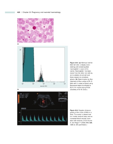
                                                                                      Figure 30.5   (a)  Kleihauer test for


                                                                            fetal red cells; a deeply eosin -
                                                                              staining cell containing fetal
                                                                            haemoglobin is seen at the
                                                                            centre. Haemoglobin has been
                                                                            eluted from the other red cells by
                                                                            an incubation at acid pH and
                                                                            these appear as colourless
                                                                            ghosts.  (b)  Determination by fl ow
                                                                            cytometry of the number of Rh D
                        10 -1       10 0       10 1       10 2       10 3   fetal cells in maternal blood using
                                                                            fl uorescent - label of antibody to
                                            Anti-D FITC
                                                                            Rh D, the mother being Rhdd.
                    (b)                                                       (Courtesy of Dr W. Erber.)


                                                                                      Figure 30.6   Doppler ultrasono-


                                                                            graphy of the circle of Willis in a
                                                                            fetus. The cursor is placed over
                                                                            the middle cerebral artery and an
                                                                            increased blood velocity corre-
                                                                            lates with anaemia.  (From Kumar
                                                                            S. and Regan F. (2005)  BMJ   330 ,
                                                                            1255 – 8, with permission.)

