
Page 62 - Essential Haematology
P. 62
48 / Chapter 3 Hypochromic anaemias
important as the source of the haemorrhage leading
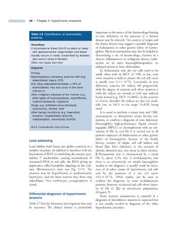
Table 3.8 Classifi cation of sideroblastic
anaemia. to iron deficiency or the presence of a chronic
disease may be revealed. The country of origin and
the family history may suggest a possible diagnosis
Hereditary
X chromosome linked ALA - S mutation or rarely of thalassaemia or other genetic defect of haemo-
with spinocerebellar degeneration and ataxia globin. Physical examination may also be helpful in
Usually occurs in males, transmitted by females; determining a site of haemorrhage, features of a
also occurs rarely in females chronic inflammatory or malignant disease, koilo-
Other rare types (see text) nychia or, in some haemoglobinopathies, an
enlarged spleen or bony deformities.
Acquired
In thalassaemia trait the red cells tend to be
Primary
small, often with an MCV of 70 fL or less, even
Myelodysplasia (refractory anaemia with ring when anaemia is mild or absent; the red cell count
sideroblasts) (see p. 215 ) 12
is usually over 5.5 × 10 /L. Conversely, in iron
N.B. Ring sideroblast formation ( < 15% of
deficiency anaemia the indices fall progressively
erythroblasts) may also occur in the bone
with the degree of anaemia and when anaemia is
marrow in:
mild the indices are normal or only just reduced
other malignant diseases of the marrow (e.g.
below normal (e.g. MCV 75 – 80 fL). In the anaemia
other types of myelodysplasia, myelofi brosis,
myeloid leukaemia, myeloma) of chronic disorders the indices are also not mark-
drugs, e.g. antituberculous (isoniazid, edly low, an MCV in the range 75 – 82 fL being
cycloserine), alcohol, lead usual.
other benign conditions (e.g. haemolytic It is usual to perform a serum iron and TIBC
anaemia, megaloblastic anaemia, measurement, or alternatively serum ferritin esti-
malabsorption, rheumatoid arthritis) mation, to confirm a diagnosis of iron defi ciency.
Haemoglobin high - performance liquid chroma-
ALA - S, δ - aminolevulinic acid synthase. tography (HPLC ) or electrophoresis with an esti-
mation of Hb A 2 and Hb F is carried out in all
patients suspected of thalassaemia or other genetic
defect of haemoglobin, because of the family
Lead p oisoning
history, country of origin, red cell indices and
Lead inhibits both haem and globin synthesis at a blood film. Iron deficiency or the anaemia of
number of points. In addition it interferes with the chronic disorders may also occur in these subjects.
breakdown of RNA by inhibiting the enzyme pyri- β - Thalassaemia trait is characterized by a raised
′
midine 5 nucleotidase, causing accumulation of Hb A 2 above 3.5%, but in α - thalassaemia trait
denatured RNA in red cells, the RNA giving an there is no abnormality on simple haemoglobin
appearance called basophilic stippling on the ordi- studies so the diagnosis is usually made by exclu-
nary (Romanowsky) stain (see Fig. 2.17 ). Th e sion of all other causes of hypochromic red cells
anaemia may be hypochromic or predominantly and by the presence of a red cell count
12
haemolytic, and the bone marrow may show ring > 5.5 × 10 /L. DNA studies can be used to
sideroblasts. Free erythrocyte protoporphyrin is confirm the diagnosis. In some α - thalassaemia
raised. patients, however, occasional red cells show depos-
its of Hb H ( β 4 ) in reticulocyte preparations
(Chapter 7 ).
Differential d iagnosis of h ypochromic
a naemia Bone marrow examination is essential if a
diagnosis of sideroblastic anaemia is suspected but
Table 3.7 lists the laboratory investigations that may is not usually needed in diagnosis of the other
be necessary. The clinical history is particularly hypochromic anaemias.