
Page 67 - Essential Haematology
P. 67
Chapter 4 Iron overload / 53
The consequent iron overload damages paren- with cardiomyopathy in children, adolescents or
chymal cells and patients may present in adult life young adults. However, ferroportin mutations
with hepatic disease, endocrine disturbances such as usually cause reticuloendothelial but not parenchy-
diabetes mellitus or impotence, melanin skin pig- mal cell iron overload but may rarely cause paren-
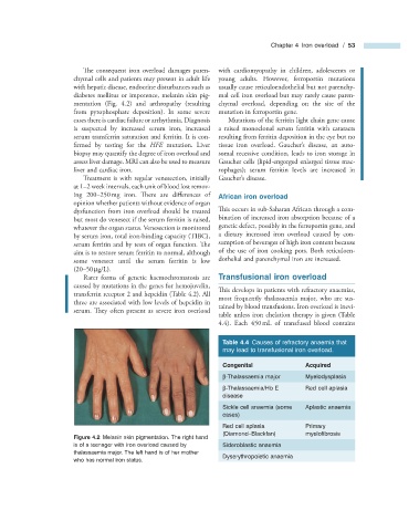
mentation (Fig. 4.2 ) and arthropathy (resulting chymal overload, depending on the site of the
from pyrophosphate deposition). In some severe mutation in ferroportin gene.
cases there is cardiac failure or arrhythmia. Diagnosis Mutations of the ferritin light chain gene cause
is suspected by increased serum iron, increased a raised monoclonal serum ferritin with cataracts
serum transferrin saturation and ferritin. It is con- resulting from ferritin deposition in the eye but no
’
firmed by testing for the HFE mutation. Liver tissue iron overload. Gaucher s disease, an auto-
biopsy may quantify the degree of iron overload and somal recessive condition, leads to iron storage in
assess liver damage. MRI can also be used to measure Gaucher cells (lipid - engorged enlarged tissue mac-
liver and cardiac iron. rophages); serum ferritin levels are increased in
Treatment is with regular venesection, initially Gaucher ’ s disease.
at 1 – 2 week intervals, each unit of blood lost remov-
ing 200 – 250 mg iron. There are diff erences of African i ron o verload
opinion whether patients without evidence of organ
dysfunction from iron overload should be treated This occurs in sub - Saharan African through a com-
but most do venesect if the serum ferritin is raised, bination of increased iron absorption because of a
whatever the organ status. Venesection is monitored genetic defect, possibly in the ferroportin gene, and
by serum iron, total iron - binding capacity (TIBC), a dietary increased iron overload caused by con-
serum ferritin and by tests of organ function. Th e sumption of beverages of high iron content because
aim is to restore serum ferritin to normal, although of the use of iron cooking pots. Both reticuloen-
some venesect until the serum ferritin is low dothelial and parenchymal iron are increased.
(20 – 50 μ g/L).
Rarer forms of genetic haemochromatosis are Transfusional i ron o verload
caused by mutations in the genes for hemojuvelin,
This develops in patients with refractory anaemias,
transferrin receptor 2 and hepcidin (Table 4.2 ). All
most frequently thalassaemia major, who are sus-
three are associated with low levels of hepcidin in
tained by blood transfusions. Iron overload is inevi-
serum. They often present as severe iron overload
table unless iron chelation therapy is given (Table
4.4 ). Each 450 mL of transfused blood contains
Table 4.4 Causes of refractory anaemia that
may lead to transfusional iron overload.
Congenital Acquired
β - Thalassaemia major Myelodysplasia
β - Thalassaemia/Hb E Red cell aplasia
disease
Sickle cell anaemia (some Aplastic anaemia
cases)
Red cell aplasia Primary
(Diamond – Blackfan) myelofi brosis
Figure 4.2 Melanin skin pigmentation. The right hand
is of a teenager with iron overload caused by Sideroblastic anaemia
thalassaemia major. The left hand is of her mother
Dyserythropoietic anaemia
who has normal iron status.