
Page 65 - Essential Haematology
P. 65
Chapter 4 Iron overload / 51
There is no physiological mechanism for eliminat- out to determine the degree of organ damage caused
ing excess iron from the body and so iron absorp- by iron (Table 4.3 ). The serum ferritin is the most
tion is normally carefully regulated to avoid widely used test and this and the percentage satura-
accumulation. Iron overload (haemosiderosis) tion of transferrin (iron - binding capacity) are useful
occurs in disorders associated with excessive absorp- screening tests for iron overload and for monitoring
tion or may result from repeated blood transfusions its treatment. Liver biopsy with staining for iron
in patients with severe chronic anaemias. Excessive and chemical analysis of iron content is useful for
iron deposition in tissues can cause serious damage assessing both parenchymal iron (hepatic cells) and
to organs (haemochromatosis), particularly the reticuloendothelial iron in Kupffer cells. Magnetic
heart, liver and endocrine organs. The causes of iron resonance imaging (MRI), particularly the T2 *
overload are listed in Table 4.1 and of genetic technique, is the best non - invasive guide to liver
haemochromatosis in Table 4.2 . and cardiac iron.
Assessment of i ron s tatus
Hereditary ( g enetic, p rimary)
The tests that can be performed to assess iron over- h aemochromatosis
load are listed in Table 4.3 . Tests may also be carried
This is a group of diseases in which there is excessive
absorption of iron from the gastrointestinal tract
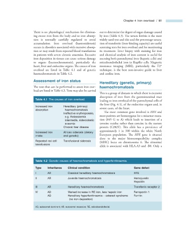
Table 4.1 The causes of iron overload.
leading to iron overload of the parenchymal cells of
the liver (Fig. 4.1 ), of the endocrine organs and, in
Increased iron Hereditary (primary)
severe cases, of the heart.
absorption haemochromatosis
The most common gene involved is HFE and
Ineffective erythropoiesis,
most patients are homozygous for a missense muta-
e.g. thalassaemia
intermedia, sideroblastic tion (845 G to A) which leads to insertion of a
anaemia tyrosine residue rather than cysteine in the mature
Chronic liver disease protein (C282Y). This allele has a prevalence of
approximately 1 in 300 within the white North
Increased iron African siderosis (dietary
European population. Th e HFE gene is situated
intake and genetic)
close to the major histocompatibility complex
Repeated red cell Transfusional siderosis
(MHC) locus on chromosome 6. Th e abnormal
transfusions
allele is associated with HLA - A3 and - B8. Only a
Table 4.2 Genetic causes of haemochromatosis and hyperferritinaemia.
Type Inheritance Clinical condition Gene defect
I AR Classical hereditary haemochromatosis HFE
II AR Juvenile haemochromatosis Hemojuvelin
Hepcidin
III AR Hereditary haemochromatosis Transferrin receptor 2
IV AD Marked increase in RE iron, less hepatic iron Ferroportin 1
AD Hereditary hyperferritinaemia – cataract syndrome Ferritin
(no iron deposition)
AD, autosomal dominant; AR, autosomal recessive; RE, reticuloendothelial.