
Page 58 - Essential Haematology
P. 58
44 / Chapter 3 Hypochromic anaemias
Serum f erritin upper and lower gastrointestinal endoscopy and/or
radiology (e.g. computed tomography (CT) of the
A small fraction of body ferritin circulates in the
pneumocolon) or virtual colonscopy using the 3D
serum, the concentration being related to tissue,
colon system (Figs 3.12 and 3.13 ). Tests for parietal
particularly reticuloendothelial, iron stores. Th e
normal range in men is higher than in women (Fig.
3.11 ). In iron deficiency anaemia the serum ferritin
is very low while a raised serum ferritin indicates
iron overload or excess release of ferritin from
damaged tissues or an acute phase response (e.g. in
infl ammation). The serum ferritin is normal or
raised in the anaemia of chronic disorders.
Investigation of the c ause of i ron
d efi ciency (Fig. 3.12 )
In premenopausal women, menorrhagia and/or
repeated pregnancies are the usual causes of the
deficiency. If these are not present other causes must
be sought. In some patients with menorrhagia a
clotting or platelet abnormality (e.g. von Willebrand
disease) is present. In men and postmenopausal
women, gastrointestinal blood loss is the main cause
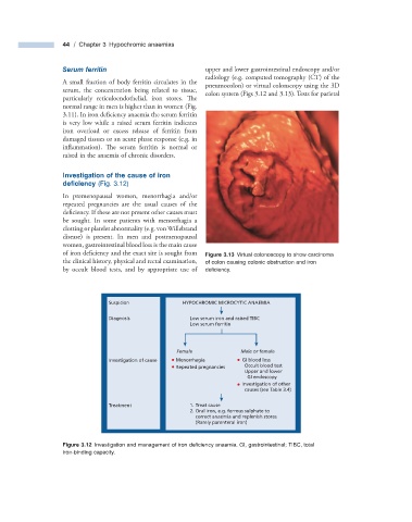
of iron deficiency and the exact site is sought from Figure 3.13 Virtual colonoscopy to show carcinoma
the clinical history, physical and rectal examination, of colon causing colonic obstruction and iron
by occult blood tests, and by appropriate use of defi ciency.
Suspicion HYPOCHROMIC MICROCYTIC ANAEMIA
Diagnosis Low serum iron and raised TIBC
Low serum ferritin
Female Male or female
Investigation of cause Menorrhagia GI blood loss
Repeated pregnancies Occult blood test
Upper and lower
GI endoscopy
Investigation of other
causes (see Table 3.4)
Treatment 1. Treat cause
2. Oral iron, e.g. ferrous sulphate to
correct anaemia and replenish stores
(Rarely parenteral iron)
Figure 3.12 Investigation and management of iron defi ciency anaemia. GI, gastrointestinal; TIBC, total
iron - binding capacity.