
Page 55 - Essential Haematology
P. 55
Chapter 3 Hypochromic anaemias / 41
(a)
(b) (c)
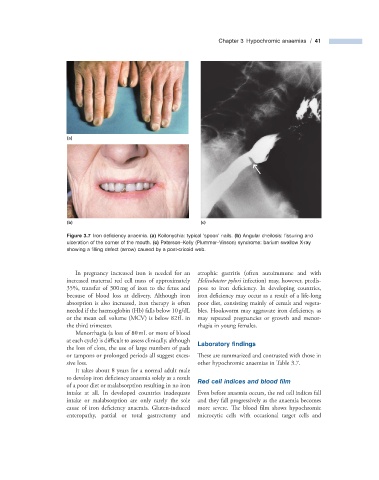
Figure 3.7 Iron defi ciency anaemia. (a) Koilonychia: typical ‘ spoon ’ nails. (b) Angular cheilosis: fi ssuring and
ulceration of the corner of the mouth. (c) Paterson – Kelly (Plummer – Vinson) syndrome: barium swallow X - ray
showing a fi lling defect (arrow) caused by a post - cricoid web.
In pregnancy increased iron is needed for an atrophic gastritis (often autoimmune and with
increased maternal red cell mass of approximately Helicobacter pylori infection) may, however, predis-
35%, transfer of 300 mg of iron to the fetus and pose to iron deficiency. In developing countries,
because of blood loss at delivery. Although iron iron deficiency may occur as a result of a life - long
absorption is also increased, iron therapy is often poor diet, consisting mainly of cereals and vegeta-
needed if the haemoglobin (Hb) falls below 10 g/dL bles. Hookworm may aggravate iron defi ciency, as
or the mean cell volume (MCV) is below 82 fL in may repeated pregnancies or growth and menor-
the third trimester. rhagia in young females.
Menorrhagia (a loss of 80 mL or more of blood
at each cycle) is difficult to assess clinically, although Laboratory fi ndings
the loss of clots, the use of large numbers of pads
or tampons or prolonged periods all suggest exces- These are summarized and contrasted with those in
sive loss. other hypochromic anaemias in Table 3.7 .
It takes about 8 years for a normal adult male
to develop iron defi ciency anaemia solely as a result Red c ell i ndices and b lood fi lm
of a poor diet or malabsorption resulting in no iron
intake at all. In developed countries inadequate Even before anaemia occurs, the red cell indices fall
intake or malabsorption are only rarely the sole and they fall progressively as the anaemia becomes
cause of iron deficiency anaemia. Gluten - induced more severe. The blood film shows hypochromic
enteropathy, partial or total gastrectomy and microcytic cells with occasional target cells and