
Page 56 - Essential Haematology
P. 56
42 / Chapter 3 Hypochromic anaemias
Table 3.4 Causes of iron defi ciency.
Chronic blood loss
Uterine
Gastrointestinal, e.g. peptic ulcer, oesophageal
varices, aspirin (or other non - steroidal anti -
infl ammatory drugs) ingestion, partial
gastrectomy, carcinoma of the stomach,
caecum, colon or rectum, hookworm,
angiodysplasia, colitis, piles, diverticulosis
Rarely, haematuria, haemoglobinuria, pulmonary
haemosiderosis, self - infl icted blood loss
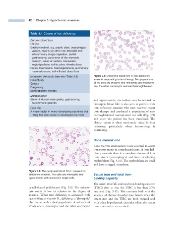
Increased demands (see also Table 3.3 ) Figure 3.9 Dimorphic blood fi lm in iron defi ciency
Prematurity anaemia responding to iron therapy. Two populations
Growth of red cells are present: one microcytic and hypochro-
Pregnancy mic, the other normocytic and well haemoglobinized.
Erythropoietin therapy
Malabsorption
Gluten - induced enteropathy, gastrectomy, and hypochromic; the indices may be normal. A
autoimmune gastritis
dimorphic blood film is also seen in patients with
Poor diet iron deficiency anaemia who have received recent
A major factor in many developing countries but iron therapy and produced a population of new
rarely the sole cause in developed countries haemoglobinized normal - sized red cells (Fig. 3.9 )
and when the patient has been transfused. Th e
platelet count is often moderately raised in iron
deficiency, particularly when haemorrhage is
continuing.
Bone m arrow i ron
Bone marrow examination is not essential to assess
iron stores except in complicated cases. In iron defi -
ciency anaemia there is a complete absence of iron
from stores (macrophages) and from developing
erythroblasts (Fig. 3.10 ). The erythroblasts are small
and have a ragged cytoplasm.
Figure 3.8 The peripheral blood fi lm in severe iron
defi ciency anaemia. The cells are microcytic and Serum i ron and t otal i ron -
hypochromic with occasional target cells. b inding c apacity
The serum iron falls and total iron - binding capacity
pencil - shaped poikilocytes (Fig. 3.8 ). Th e reticulo- (TIBC) rises so that the TIBC is less than 20%
cyte count is low in relation to the degree of saturated (Fig. 3.11 ). Th is contrasts both with the
anaemia. When iron deficiency is associated with anaemia of chronic disorders (see below) when the
severe folate or vitamin B 12 deficiency a ‘ dimorphic ’ serum iron and the TIBC are both reduced and
film occurs with a dual population of red cells of with other hypochromic anaemias where the serum
which one is macrocytic and the other microcytic iron is normal or even raised.