
Page 57 - Essential Haematology
P. 57
Chapter 3 Hypochromic anaemias / 43
(a) (b)
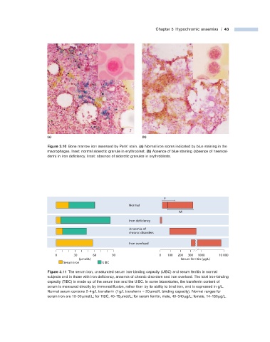
Figure 3.10 Bone marrow iron assessed by Perls ’ stain. (a) Normal iron stores indicated by blue staining in the
macrophages. Inset: normal siderotic granule in erythroblast. (b) Absence of blue staining (absence of haemosi-
derin) in iron defi ciency. Inset: absence of siderotic granules in erythroblasts.
F
Normal
M
Iron deficiency
Anaemia of
chronic disorders
Iron overload
0 30 60 90 0 100 200 300 1000 10 000
(μmol/L) Serum ferritin (μg/L)
Serum iron UIBC
Figure 3.11 The serum iron, unsaturated serum iron - binding capacity (UIBC) and serum ferritin in normal
subjects and in those with iron defi ciency, anaemia of chronic disorders and iron overload. The total iron - binding
capacity (TIBC) is made up of the serum iron and the UIBC. In some laboratories, the transferrin content of
serum is measured directly by immunodiffusion, rather than by its ability to bind iron, and is expressed in g/L.
Normal serum contains 2 – 4 g/L transferrin (1 g/L transferrin = 20 μ mol/L binding capacity). Normal ranges for
serum iron are 10 – 30 μ mol/L; for TIBC, 40 – 75 μ mol/L; for serum ferritin, male, 40 – 340 μ g/L; female, 14 – 150 μ g/L.