
Page 80 - Essential Haematology
P. 80
66 / Chapter 5 Macrocytic anaemias
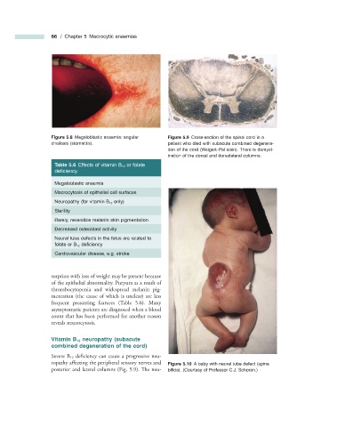
Figure 5.8 Megaloblastic anaemia: angular Figure 5.9 Cross - section of the spinal cord in a
cheilosis (stomatitis). patient who died with subacute combined degenera-
tion of the cord (Weigert – Pal stain). There is demyel-
ination of the dorsal and dorsolateral columns.
Table 5.6 Effects of vitamin B 12 or folate
defi ciency.
Megaloblastic anaemia
Macrocytosis of epithelial cell surfaces
Neuropathy (for vitamin B 12 only)
Sterility
Rarely, reversible melanin skin pigmentation
Decreased osteoblast activity
Neural tube defects in the fetus are related to
folate or B 12 defi ciency
Cardiovascular disease, e.g. stroke
sorption with loss of weight may be present because
of the epithelial abnormality. Purpura as a result of
thrombocytopenia and widespread melanin pig-
mentation (the cause of which is unclear) are less
frequent presenting features (Table 5.6 ). Many
asymptomatic patients are diagnosed when a blood
count that has been performed for another reason
reveals macrocytosis.
Vitamin B 12 n europathy ( s ubacute
c ombined d egeneration of the c ord)
Severe B 12 deficiency can cause a progressive neu-
ropathy affecting the peripheral sensory nerves and Figure 5.10 A baby with neural tube defect (spina
posterior and lateral columns (Fig. 5.9 ). Th e neu- bifi da). (Courtesy of Professor C.J. Schorah.)