
Page 216 - parasitology for medical and clinical laboratoryprofessionals
P. 216
196 CHAPTER 8
include the initial inflammatory reaction at the site where In the next larval stage, the cercariae produced in
the metacercariae penetrate the skin and is commonly the snail are released into the water to infect new hosts.
called swimmer’s itch. Abdominal pain and weight These cercariae contaminate the water and penetrate the
loss are common and bloody diarrhea may occur along skin of humans exposed to the water. Following entry
with eosinophilia and hepatosplenomegaly (enlarge- through the skin of the humans, the cercariae lose their
ment of the liver and spleen). Painful urination with the tails and become known as schistosomula. These tailless
excretion of bloody urine may develop in infections by forms enter the blood circulation (hence the name blood
S. hematobium. flukes) and migrate through the body until they reach
their final position in blood vessels, where they mature
Life Cycle into the metacercariae that are encysted in the host.
They inhabit the blood vessels near the intestinal tract
The adult worms live in blood vessels associated with and the liver in the cases of S. mansoni and S. japonicum,
the intestine or bladder depending upon the species. and near the urinary bladder for S. hematobium. At this
After the male and female worms mate, the ova produced point they are now ready to leave the body in either urine
migrate to the intestine or to the lumen of the urinary (S. hematobium) or in feces (S. mansoni and
bladder and the females produce eggs that are passed M. japonicum) by being discharged into the water.
out with feces or urine. When the eggs of S. hematobium
enter the water, the larval stages of the organism, called Disease Transmission
miracidia, are released from the eggs. These potentially
invasive forms then begin the search for a suitable snail Humans enter the water where snails have become
host. When they find this intermediate host, the snail, infected by the miracidia of one species of Schistosoma.
they bore into the tissues of the snail and a period of The miracidia, which develop into cercariae, are released
multiplication commences. from the snail and penetrate the skin of the human host.
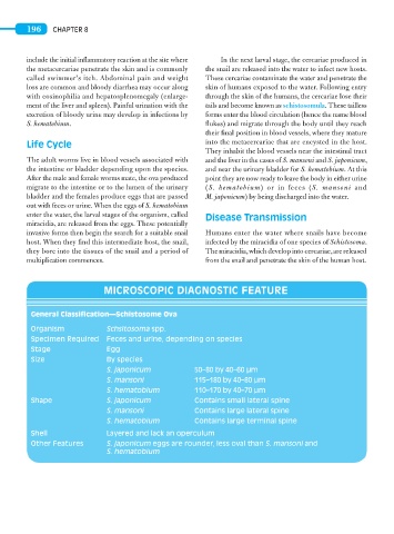
MICROSCOPIC DIAGNOSTIC FEATURE
General Classification—Schistosome Ova
Organism Schsitosoma spp.
Specimen Required Feces and urine, depending on species
Stage Egg
Size By species
S. japonicum 50–80 by 40–60 μm
S. mansoni 115–180 by 40–80 μm
S. hematobium 110–170 by 40–70 μm
Shape S. japonicum Contains small lateral spine
S. mansoni Contains large lateral spine
S. hematobium Contains large terminal spine
Shell Layered and lack an operculum
Other Features S. japonicum eggs are rounder, less oval than S. mansoni and
S. hematobium