
Page 147 - DUOKOPT BIBLIOBOOK
P. 147
EFFICACY
Acta Ophthalmologica 2010
Table 4. Continued increase in arterial pCO 2 – a stimulus
that produces extracellular acidosis –
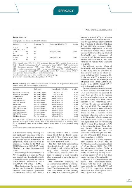
Demographic and clinical variables (146 patients)
also strongly increases choroidal blood
Variables n Progressed, % Univariate HR (95% CI) p-value flow (Friedman & Chandra 1972; Riva
& Petrig 1995; Schmetterer et al. 1996).
< 12.8 40 38 Nevertheless, experiments in isolated
EDV CRA, cm ⁄ s pre-contracted bovine retinal arteries
< 4.3 102 37 1.01 (0.64–1.60) 0.949
‡ 4.3 113 36 indicate that the vasodilatory effects of
RI CRA, cm ⁄ s dorzolamide are independent of
< 0.66 123 33 0.87 (0.54–1.36) 0.512 changes in pH because dorzolamide-
‡ 0.66 92 41 induced vasodilatation is also seen
when the pH remains stable (Josefsson
HR = hazard ratio; 95% CI = 95% confidence interval; SBP = systolic blood pressure; et al. 2004).
DBP = diastolic blood pressure; MAP = mean arterial pressure; HBP = high blood pressure;
IOP = intraocular pressure; MD = mean defect; PSD = pattern standard deviation; The different vascular effects of
OPP = ocular perfusion pressure; CCT = central corneal thickness; OA = ophthalmic artery; dorzolamide and brinzolamide found
PSV = peak systolic velocity; EDV = end-diastolic velocity; RI = resistivity index; SPCA = in this study may be explained by
short posterior ciliary artery; CRA = central retinal artery. their different abilities to inhibit car-
For all variables, the second listed variable is the reference group. bonic anhydrase (CA) isoenzyme IV.
p-values were considered statistically significant at < 0.05. In the eye, CA-IV is located in the
endothelial cells of the choriocapillaris
(Hageman et al. 1991). Additionally,
Table 5. Follow-up potential risk factors associated with visual field progression in a univariate Sender et al. (1994) found CA-IV
analysis among 146 patients included in the study. activity in muscle capillaries.
The vasorelaxation observed in vitro
Variable Reference Hazard ratio (95% CI) p-value
or after systemic administration of
Mean IOP at follow-up Per mmHg higher 1.19 (1.01–1.37) 0.038 CAIs can therefore be assumed to
Mean IOP fluctuation Per mmHg higher 1.12 (1.02–1.21) 0.013 result from an effect on the vascular
IOP reduction Per mmHg lower 0.88 (0.73–1.04) 0.103 smooth muscle cells alone or on these
OPP increase Per mmHg higher 0.86 (0.73–1.02) 0.097
PSV OA increase Per cm ⁄ s higher 0.87 (0.54–1.35) 0.498 cells in interplay with other cellular
EDV OA increase Per cm ⁄ s higher 0.52 (0.40–0.70) < 0.001 elements in the surrounding tissue.
RI decrease Per 0.01 unit lower 0.55 (0.39–0.82) 0.001 However, this response depended on
PSV CRA increase Per cm ⁄ s higher 1.03 (0.66–1.63) 0.879 the type of CAI. The vasorelaxing
EDV CRA increase Per cm ⁄ s higher 0.91 (0.57–1.43) 0.664 effect of dorzolamide was significantly
RI CRA decrease Per 0.01 unit lower 0.86 (0.57–1.18) 0.206 reduced in isolated arterioles, as evi-
PSV SPCA increase Per cm ⁄ s higher 0.92 (0.58–1.43) 0.683 denced by both a reduction in the
EDV SPCA increase Per cm ⁄ s higher 0.63 (0.42–0.90) 0.004
RI SPCA decrease Per 0.01 unit lower 0.58 (0.41–0.88) 0.002 maximum relaxation and an increase
in EC50 (median effective concentra-
95% CI = 95% confidence interval; IOP = intraocular pressure; OPP = ocular perfusion tion) (Kehler et al. 2007). It is there-
pressure; PSV = peak systolic velocity; EDV = end-diastolic velocity; RI = resistivity index; fore highly likely that the vasorelaxing
OA = ophthalmic artery; CRA = central retinal artery; SPCA = short posterior ciliary effects of these drugs depend on CAs
artery. in the perivascular tissue. These effects
p-values were considered statistically significant at < 0.05.
may involve different membrane-
bound isoenzymes IV and XIV
IOP fluctuation during follow-up was increasing evidence that a reduced located in retinal astrocytes and Mu ¨ l-
not significantly associated with pro- ocular blood flow is directly associ- ler cells (Wistrand 2000; Inoue et al.
gression. Our study found no relation- ated with VF loss (Galassi et al. 2003; 2004; Nagelhus et al. 2005).
ship between age and worsening of VF Satilmis et al. 2003; Martinez & San- Dorzolamide is a potent sulphona-
progression, which contrasts with the chez 2005; Zeitz et al. 2006). mide inhibitor of CA-IV, with an
conclusions reached by the AGIS and The fact that both combinations IC50 (median inhibition concentration)
EMGT (Leske et al. 2003, 2007; Nouri- (dorzolamide–timolol and brinzola- of 6.9 nm (Sugrue 2000). The IC50
Mahdavi et al. 2004a; Bengtsson et al. mide–timolol) had a similar IOP-low- for brinzolamide against CA-IV is
2007). ering effect, although different 45.3 nm (Stams et al. 1998). In other
We found significantly lower rates of vascular effects, provides further evi- words, the inhibitory activity of dor-
VF progression at 5 years in POAG dence to support a local vasoactive zolamide against CA-IV is 6.5 times
eyes treated with dorzolamide–timolol effect as opposed to an ocular tension greater than that of brinzolamide.
compared with eyes treated with brinzo- mechanism. The rates of VF progression in eyes
lamide–timolol (multivariate HR = The mechanism underlying the treated with dorzolamide–timolol,
0.65, 95% CI 0.41–0.90; p = 0.009). vasodilatory effect of CAIs in retrobul- detected with the event- and trend-
These findings may be explained by bar vessels has not been fully estab- based methods, were 24% (24⁄ 99) and
the fact that dorzolamide significantly lished. There is evidence that CAIs may 27% (27⁄ 99), respectively, substan-
increases haemodynamic parameters cause extracellular acidosis (Friberg tially lower than rates reported in the
in retrobulbar vessels and there is et al. 1990). It is worth noting that an EMGT (Leske et al. 2003).
549 147