
Page 36 - Live-cellanalysis handbook
P. 36
Live-Cell Analysis Handbook — Third Edition
Assessment of immune cell killing of cancer cells using caspase signaling
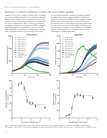
Capsase-3/7 was used to evaluate targeted tumor cell death in A concentration-dependent decrease in proliferation (IC50
a co-culture Antibody-Dependent Cell-mediated Cytotoxicity 8.1 ng/mL) and increase in apoptosis (IC50 4.6 ng/mL) was
(ADCC) assay to measure tumor cell apoptosis and for visual measured in Her2-positive SKOV3 cells. No response was seen
validation of tumor-immune cell interactions. To demonstrate in Her2-negative A549 cells (data not shown). These data
the utility of this approach, ADCC cell death was measured demonstrate that live cell imaging can be used to discern the
via Caspase-3/7 induction in Her2 positive SKOV3 or negative full-time course and specificity of immune cell killing, which
A549 NucLight Red cells (1.6K/well) co-cultured with with are traditionally conducted as flow cytometry or biochemical
PBMCs (8K/well) in the presence of trastuzumab, a clinically readouts, requiring selection of end points and lack visual
used monoclonal antibody for Her-2 positive sold cancers. confirmation of cellular interactions.
Proliferation Apoptosis
Figure 7. Trastuzumab induced ADCC in Her2 positive SKOV3 cells. SK-OV-3 cancer cells were seeded in combination with PBMCs in the presence of
trastuzumab to induce ADCC. A concentration-dependent decrease in proliferation (IC50 8.1 ng/mL) and an increase in apoptosis (IC50 4.6 ng/mL) was seen.
34