
Page 39 - Live-cellanalysis handbook
P. 39
Kinetic Cytotoxicity Assays
Shortcomings of Traditional Assays Live-Cell Imaging and Analysis Approaches
• Assays result in a single, user-defined time • The assay provides a full kinetic readout of cytotoxicity over multiple days, eliminating
point measurement. the need for determining a single, optimum, assay endpoint a-priori which can vary
considerably for different cell types and for different compound treatment conditions.
• Lack of ability to assess biological activity
over time limits predictive nature. • Addition of IncuCyte CytoTox reagents to normal, healthy cells are nonperturbing to
cell growth and morphology.
• Manipulations can result in the loss of cells
or critical data in experiments where • Cells can be simultaneously labeled with an IncuCyte CytoTox reagent and NucLight
cells undergo cell death at different rates nuclear labeling reagent to measure cytotoxicity and cell proliferation.
according to treatment conditions.
• 96- and 384-well assay format follows a homogeneous “mix and read” protocol which can
be run over multiple days in full media. No wash or lifting steps required, negating the
concern that cells are lost during the experiment or labeling process.
• All data points and temporal data curves can be validated by individual images or time-
lapse movies respectively. The kinetic readout of the IncuCyte® system provides both
high definition (HD) phase as well as quantitative fluorescent imaging.
Sample Results
Quantitative measurement of cytotoxicity using IncuCyte CytoTox reagent
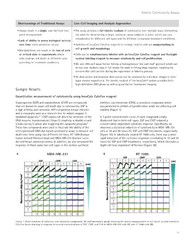
Staurosporine (SSP) and camptothecin (CMP) are compounds inhibitor, cycloheximide (CHX), a cytostatic compound which
that are known to cause cell death due to cytotoxicity. SSP is was predicted to inhibit cell proliferation while not affecting cell
a high affinity, non-selective, ATP-competitive kinase inhibitor viability (Figure 1).
and is classically used as a research tool to induce caspase-3
mediated apoptosis. CMP causes cell death by inhibition of the A 7-point concentration curve of each compound clearly
6, 7
DNA enzyme, topoisomerase I (topo I), resulting in double strand illustrated that in both cell types, SSP and CMP induced a
breaks during S-phase and triggering the apoptotic program. concentration-dependent cytotoxic response. Specifically, we
8
These two compounds were used to illustrate the ability of the observed a statistical induction of cytotoxicity in MDA-MB-231
cell impermeant DNA dye based cytotoxicity assay to measure cell cells at 16 and 26 hours for SSP and CMP treatments, respectively
death over-time using two different cell lines, HT 1080 (human (Figure 1A). In identically treated HT 1080 cells, there was a more
tumor derived fibrosarcoma) and MDA-MB-231 (human tumor rapid induction of the cytotoxic responses correlating to 12 and 22
derived breast adenocarcinoma. In addition, we also measured the hours for SSP and CMP treatments, respectively, which illustrates a
response of these same two cell types to the protein synthesis slight cell type dependent difference (Figure 1B).
A MDA-MB-231 B HT 1080
Figure 1. Discrimination of cytotoxic and cytostatic compounds. 96-well microplate graph showing the kinetic measurement of cell death as determined by
CytoTox Green staining in response to several concentrations of SSP, CMP, and CHX in MDA-MB-231 cells (A) and HT 1080 cells (B).
37