
Page 309 - Zoo Animal Learning and Training
P. 309
Chapter 15 Dental Skills for the Veterinary Assistant 293
Intraoral Radiography member working in dental radiology must follow as it
orients the veterinarian when reading a dental film.
More and more veterinary clinics are understanding
Dental radiographs are an essential part of each and the value of dental radiographs and in an effort to reduce
every oral examination. The crown is just the “tip of the anesthesia time are turning to digital dental radiograph
iceberg,” so to speak. Approximately 42% of dental sensors. These sensors allow the radiograph image to
pathology is found subgingivally. Radiographs will help appear on a computer monitor within seconds of
diagnose pathology that is not visible from the surface, shooting.
confirm suspect pathology, as well as help demonstrate There are two types of digital systems used to produce
the pathology to the client. Survey radiographs can also a dental radiograph. Digital radiography (DL) uses a
increase your clinic’s revenue. sensor that is inside a rigid plastic wafer. The wafer is
Dental radiography is part of the diagnostic workup
for dental disease. With the exception of positioning, placed inside the patient’s mouth and then exposed.
The wafer is connected to the computer by a wire or is
dental radiography follows the same principles as gen- wireless and transfers the image to the computer. The
eral radiology, including the use of PPE (see Chapter 16). wafer comes in two sizes and can be damaged if dropped.
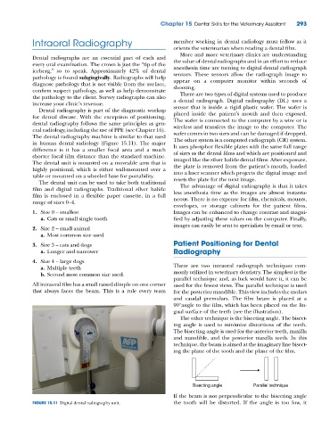
The dental radiography machine is similar to that used The other system is a computed radiograph (CR) system.
in human dental radiology (Figure 15.11). The major It uses phosphor flexible plates with the same full range
difference is it has a smaller focal area and a much of sizes as the dental films and which are positioned and
shorter focal film distance than the standard machine. imaged like the silver halide dental films. After exposure,
The dental unit is mounted on a moveable arm that is the plate is removed from the patient’s mouth, loaded
highly positional, which is either wall‐mounted over a into a laser scanner which projects the digital image and
table or mounted on a wheeled base for portability. resets the plate for the next image.
The dental unit can be used to take both traditional
The advantage of digital radiography is that it takes
film and digital radiographs. Traditional silver halide less anesthesia time as the images are almost instanta-
film is enclosed in a flexible paper cassette, in a full neous. There is no expense for film, chemicals, mounts,
range of sizes 0–4.
envelopes, or storage cabinets for the patient films.
1. Size 0 – smallest Images can be enhanced to change contrast and magni-
a. Cats or small single tooth fied by adjusting these values on the computer. Finally,
images can easily be sent to specialists by email or text.
2. Size 2 – small animal
a. Most common size used
3. Size 3 – cats and dogs Patient Positioning for Dental
a. Longer and narrower Radiography
4. Size 4 – large dogs
a. Multiple teeth There are two intraoral radiograph techniques com-
b. Second most common size used. monly utilized in veterinary dentistry. The simplest is the
parallel technique and, as luck would have it, it can be
All intraoral film has a small raised dimple on one corner used for the fewest views. The parallel technique is used
that always faces the beam. This is a rule every team for the posterior mandible. This view includes the molars
and caudal premolars. The film beam is placed at a
90°angle to the film, which has been placed on the lin-
gual surface of the teeth (see the illustration).
The other technique is the bisecting angle. The bisect-
ing angle is used to minimize distortions of the teeth.
The bisecting angle is used for the anterior teeth, maxilla
and mandible, and the posterior maxilla teeth. In this
technique, the beam is aimed at the imaginary line bisect-
ing the plane of the tooth and the plane of the film.
Bisecting angle Parallel technique
If the beam is not perpendicular to the bisecting angle
FIGURE 15.11 Digital dental radiography unit. the tooth will be distorted. If the angle is too low, it