Page 315 - Zoo Animal Learning and Training
P. 315
Chapter 16  Diagnostic Imaging and Endoscopy    299


                                                                   The National Committee on Radiation Protection
               Reflection                                       and Measurements (NCRP) recommends using “as low
                                                                as reasonably achievable” (ALARA) exposures when tak-
               Compare and contrast the different diagnostic    ing radiographs. This means that the personnel taking
                imaging systems.                                radiographs need to know what they are doing so the
                                                                image is right the first time. For every radiograph taken
                                                                there is more exposure to radiation increasing the
             Quality Assurance                                  chance of damage to the personnel over a lifetime. Some
                                                                states actually prohibit manual restraint of animals dur-
                                                                ing radiographic procedures. Patients are anesthetized
             The usefulness of a radiograph is limited by the quality   and positioning aids are used. This is a sensible guide-
             of the image itself. It is the veterinary technician and the   line even when not required by law. Another option is to
             assistant who actually take the radiographs, so their   rotate staff conducting radiographic procedures, espe-
             ability to produce quality films is essential. A quality film   cially in high volume practices.
             has subtle shades of white to black contrasts depending   Major safety measures are used to reduce the amount of
             on the density of the body parts. For example, a leg   radiation exposure to personnel. The following guidelines
             bone is mostly white with shades of gray because of its   must be followed to ensure a safe working environment:
             different densities, whereas muscle tissue is a fairly uni-
             form dark shade of gray because it is less dense and   1.  Each person involved in the radiograph must wear
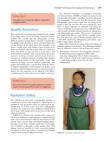
             fairly uniform in density. This contrast allows the veteri-  personal protection equipment (PPE) while
             narian to see body structures and determine if there is   exposing radiographs. This includes a lead apron
             an injury or condition present. A quality film requires   that covers the front of the body from the shoulders
             accurate measurement of the body part, correct film   to mid‐thigh, goggles, and a thyroid collar
             exposure settings, accurate patient positioning, and   (Figure 16.1).
             proper developing of the exposed film. With the advent
             of digital radiography, measurement and proper posi-
             tioning are still very important but, because the image is
             digital, the film exposure can be adjusted to be lighter
             or darker and of course there are no “films” to develop.


               Reflection

               Discuss the reason for quality assurance and the
               issues it would cause if films were not diagnostic.


             Radiation Safety


             The radiation produced by a radiography machine is
             considered  a  hazard  and  regulations  regarding  use  of
             equipment and personnel safety are governed by the
             Department of Health at the state level. Radiography
             machines are registered with a Radiation Safety Board
             which inspects the equipment to make sure it is operating
             within parameters.
               Radiation is odorless and colorless and it cannot be
             felt or seen. Radiation causes damage to rapidly dividing
             cells, such as those found in unborn and immature indi-
             viduals, bone marrow, skin, and blood‐forming tissues.
             Damage occurs when multiple small doses accumulate
             over a lifetime or through a single massive dose. This
             damage may not manifest for years or genetic damage
             can appear in future generations. Consistently following
             proper safety measures will prevent harm.          FIGURE 16.1  Lead apron and thyroid collar.

