Page 316 - Zoo Animal Learning and Training
P. 316
300    Tasks for the Veterinary Assistant


                                                                 there are breaks in the lead. Notify the inventory
                                                                 manager so that piece of equipment can be
                                                                 replaced. The assistant can help co‐workers by
                                                                 making sure this gets done!
                                                              6.  Individuals under 18 years of age and pregnant
                                                                 women are never allowed in the room when
                                                                 radiographs are being exposed.
                                                              7.  Never enter a room denoted by a radiation hazard
                                                                 symbol unless invited or it is known that a radiograph
                                                                 is not being taken at that time. Close and if possible
                                                                 lock the door to the radiation room, or indicate
                                                                 radiographs are being taken by turning on a
                                                                 warning light. This symbol denotes radiation is
                                                                 used in that location
                                                              8.  Reduce personal exposure by:
          FIGURE 16.2  Lead gloves.                              •  Staying out of the primary beam which comes
                                                                  from the X‐ray tube housing and points down at
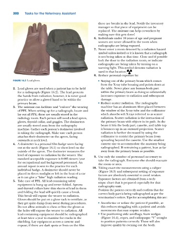
          2.  Lead gloves are used when a patient has to be held   the table. Never place any human body part
             for a radiograph (Figure 16.2). The lead protects    within the primary beam as doing so substantially
             the hands from radiation; however, it is never good   increases exposure to radiation and subsequent
             practice to allow a gloved hand to be within the     damage.
             primary beam.                                       •  Reduce scatter radiation. The radiography
          3.  The assistant can facilitate and “enforce” the wearing   machine has an aluminum filter placed between
             of PPE. When setting up for a radiograph, locate and   the window of the X‐ray tube and the collimator
             lay out all PPE; these are usually stored in the     which absorbs soft X‐rays referred to as scatter
             radiology room. Each person will need a lead apron,   radiation. Scatter radiation is the interaction of
             gloves, thyroid collar, and goggles. The dosimeters   the primary beam with objects in its path. As the
             are usually stored away from the radiography         beam it hits the body part, cassette, and table top
             machine. Gather each person’s dosimeter involved     it bounces up in an outward projection. Scatter
             in taking the radiograph. Make sure each person      radiation is further decreased by using the
             attaches their dosimeter on the apron, facing        collimator to restrict the primary beam from
             outwards at neck level.                              spreading beyond the cassette. Use the smallest
          4.  A dosimeter is a personal film badge worn facing    cassette size to accommodate the anatomy being
             out at the neck (Figure 16.1) or chest level on the   radiographed. If restraining a patient, lean as far
             outside of the apron. The dosimeter measures the     away from the primary beam as possible.
             level of exposure to radiation by the wearer. The   9.  Use only the number of personnel necessary to
             standard acceptable exposure is 0.005 sievert/year   take the radiograph. Everyone else should evacuate
             for occupational and background personnel. An       the room or area.
             annual report is sent to the practice for each   10.  Taking exact site measurements using a caliper
             individual badge. A dosimeter should never be       (Figure 16.3) and subsequent setting of exposure
             placed in direct sunlight or left in the heat of a car   factors are absolutely essential to avoid retakes.
             as it can give a “false” high radiation reading.
          5.  Take care of PPE. All lead‐containing safety       Exposure factors are obtained from a tech-
                                                                 nique chart that is prepared especially for that
               equipment is hung up and never folded. Aprons     radiography unit.
             and thyroid collars have thin sheets of lead in them   Position the patient correctly and confirm that the
             and folding the lead will quickly cause a break.    correct body part is being radiographed as per the
             This break will expose the wearer to radiation.     veterinarian’s orders. Tips for accomplishing this are:
             Gloves should be put on a glove rack to ventilate, as
             they get quite damp from sweat during procedures.   •  Anesthetize or sedate the patient if possible, as
             Do not allow animals to chew or bite the gloves as   this reduces struggling with a patient and avoids
             the holes will expose the wearer to radiation. All   movements that may require retakes.
             lead‐containing equipment should be radiographed    •  Use positioning aids: sandbags, foam wedges
             at least twice a year to examine for cracks in the   (Figure 16.4), ropes, and radiopaque “V” troughs
             shielding. Lay equipment across a cassette and       to position patients correctly. These will also
             expose; if there are dark spots or lines on the film   improve quality by evening out the body.

