
Page 317 - Zoo Animal Learning and Training
P. 317
Chapter 16 Diagnostic Imaging and Endoscopy 301
• Proper mixing of processor chemicals and
maintenance of automatic processor.
• Annual field service by a qualified representative
including machine calibration.
Strict adherence to these recommendations reduces
radiation exposure and therefore possible damage.
Once the radiation damage occurs, it is not reversible.
Radiography Abbreviations
The American Committee of Veterinary Radiologists
and Anatomists (ACVRA) is the source of anatomic and
directional terms and abbreviations used in veterinary
radiology. Understanding these abbreviations is impor-
tant because they are used to indicate what body part to
radiograph and to fill out the identification label and
FIGURE 16.3 Calipers.
log. The first letter in an abbreviation represents where
the X‐ray beam enters the body. The second letter rep-
resents where the beam exits the body. Adding these
abbreviations to your reference book is a good idea.
The following are some of the most common direc-
tional terms and abbreviations:
Left (Lt): a patient’s left side or limb
Right (Rt): a patient’s right side or limb
Dorsal (D): the upper parts of the body. This includes the
top of the head, neck, back, and tail.
Ventral (V): the lower parts of the body. This includes the
lower part of the head and neck, chest and abdomen,
and tail.
Anterior/posterior (AP): beam directed at the limb from
front to back
Palmar (Pa): the forelimb from the carpal joint distally.
Used instead of the term caudal.
Plantar (Pl): the hind limb from the tarsal joint distally.
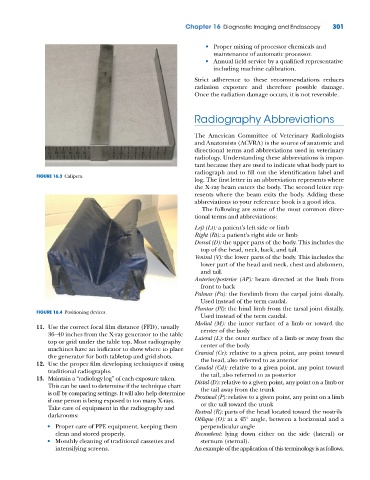
FIGURE 16.4 Positioning devices.
Used instead of the term caudal.
Medial (M): the inner surface of a limb or toward the
11. Use the correct focal film distance (FFD), usually center of the body
36–40 inches from the X‐ray generator to the table Lateral (L): the outer surface of a limb or away from the
top or grid under the table top. Most radiography center of the body
machines have an indicator to show where to place Cranial (Cr): relative to a given point, any point toward
the generator for both tabletop and grid shots.
the head, also referred to as anterior
12. Use the proper film developing techniques if using Caudal (Cd): relative to a given point, any point toward
traditional radiographs.
the tail, also referred to as posterior
13. Maintain a “radiology log” of each exposure taken. Distal (D): relative to a given point, any point on a limb or
This can be used to determine if the technique chart the tail away from the trunk
is off by comparing settings. It will also help determine Proximal (P): relative to a given point, any point on a limb
if one person is being exposed to too many X‐rays. or the tail toward the trunk
Take care of equipment in the radiography and Rostral (R): parts of the head located toward the nostrils
darkrooms:
Oblique (O): at a 45° angle, between a horizontal and a
• Proper care of PPE equipment, keeping them perpendicular angle
clean and stored properly. Recumbent: lying down either on the side (lateral) or
• Monthly cleaning of traditional cassettes and sternum (sternal).
intensifying screens. An example of the application of this terminology is as follows.