
Page 319 - Zoo Animal Learning and Training
P. 319
Chapter 16 Diagnostic Imaging and Endoscopy 303
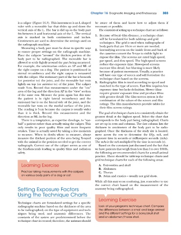
is a caliper (Figure 16.3). This instrument is an L‐shaped be aware of them and know how to adjust them if
ruler with a moveable bar that slides up and down the necessary or possible.
vertical axis of the L. The bar is slid up so the body part The constants of setting up a technique chart are as follows:
fits between it and horizontal axis of the L. The vertical 1. Because of focal–film distance, a technique chart
axis is marked in both centimeters and inches. will be formulated for both tabletop and grid
Centimeters are used to determine the settings used on techniques. The grid is used when radiographs of
the radiograph machine. body parts that are 10 cm or more are needed.
Measuring a body part must be done in specific ways
to ensure proper settings on the radiograph machine. 2. Intensifying screens on the inside front and back of
the cassettes convert the X‐rays to visible light to
Place the calipers “around” the thickest portion of the expose the film. The screens are rated high speed,
body part to be radiographed. The moveable bar is par speed, and slow speed. The high‐speed screens
allowed to settle lightly around the part being measured. reduce the exposure time. Slow‐speed screens
For example, the veterinarian orders an AP and ML of increase film detail, but blurring is more likely
the right carpus on a dog. The patient is positioned in because of patient movement. Usually, a practice
sternal recumbency and the right carpus is measured will have one type of screen and will formulate the
with the caliper. The stationary part of the bar is beneath technique chart based on the screens.
(or posterior to) the joint, and the moveable bar rests 3. Radiographic film is rated according to speed and
lightly on top (or anterior to) of the joint. The caliper matched to the type of screen. Fast film requires less
reads 3 cm. Record that measurement under the “cm” exposure time but lacks definition. Slower films
area of the log and the direction AP in the “view” section require greater exposure time and produce films
of the same row. Measure the joint again but this time with greater detail. The film–screen system is the
the patient is in right lateral recumbency and the combination of the values of the screen and film
stationary bar is on the lateral side of the joint, and the ratings. The film manufacturers provide tables for
moveable bar rests on the medial surface of the joint. their film–screen ratings.
The reading is 5 cm because the carpus is usually wider
than it is thick. Record this measurement and the The goal of technique charts is to consistently obtain the
direction as ML in the log. greatest detail at the highest speed. Select the chart that
There is a temptation, as expertise develops, to “eye- corresponds to the body part being radiographed. Charts
ball” a patient rather than actually using the caliper. This are set up in rows and columns. The first column lists the
only results in poor quality films and more frequent thickness in centimeters of the body part to be radio-
retakes. Time is actually saved by taking a few moments graphed. Once the thickness of the study site is located,
to measure. When in doubt where to measure, always move across the row to determine the kVp, mA, and
measure the thickest portion of the area being X‐rayed exposure time in seconds or milliampere seconds (mAs).
with the animal in the position needed to get the correct The mAs is the mA multiplied by the time in seconds (s).
radiograph. Correct use of the caliper serves as one of Based on the constants just discussed and the fact that
the fundamentals leading to quality films and radiation we have patients that weigh from less than l to over 100 lb,
safety. the following are recommended charts for a small animal
practice. There should be tabletop technique charts and
grid technique charts for each of the following areas:
Learning Exercise A. Extremities and skull
B. Abdomen
Practice taking measurements with the calipers C. Thorax
on various body parts of a dog or cat. D. Avian and exotics – usually not grid shots.
While all this may seem confusing, just remember to use
the correct chart based on the measurement of the
Setting Exposure Factors anatomy being radiographed.
Using the Technique Charts
Learning Exercise
Technique charts are formulated settings for a specific
radiography machine based on the thickness of the area Look at your programs technique chart. Compare
to be radiographed, on the type of equipment and tech- the differences between a small and large animal
niques being used, and anatomic differences. The and the different settings for a bone/skull shot
constants of the system are predetermined before the and an abdomen/thorax shot.
technique chart is created; therefore, the assistant should