Page 95 - Zoo Animal Learning and Training
P. 95

Chapter 5  Anatomy and Physiology    79


                                                    Epaxial
                                           Gluteal muscles                Trapezius


                                                         Fascia   Latissimus
                                              Semitendinosus        dorsi   Deltoid Biceps


                                                 Biceps
                                                  femoris

                                                                     Triceps
                             Gastrocnemius


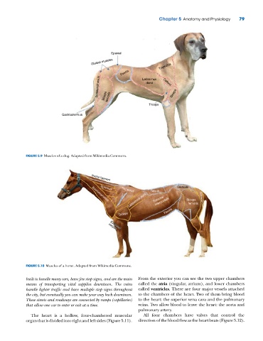
             FIGURE 5.9  Muscles of a dog. Adapted from Wikimedia Commons.


                                           Nuchal ligament
                                                                                  Gluteals
                                               Splenius
                                                                      Fascia
                                                         Trapezius
                                                            Latissimus dorsi  External  Biceps  Semitendinosus
                                                                        intercostals
                                           Brachiocephalicus
                                                                                         femoris
                                                        Triceps              Fascia


                                                    Extensor
                                                   carpus


             FIGURE 5.10  Muscles of a horse. Adapted from Wikimedia Commons.

             built to handle many cars, have few stop signs, and are the main   From the exterior you can see the two upper chambers
             means of transporting vital supplies downtown. The veins   called the atria (singular, atrium), and lower chambers
             handle lighter traffic and have multiple stop signs throughout   called ventricles. There are four major vessels attached
             the city, but eventually you can make your way back downtown.   to the chambers of the heart. Two of them bring blood
             These streets and roadways are connected by ramps (capillaries)   to the heart: the superior vena cava and the pulmonary
             that allow one car to enter or exit at a time.     veins. Two allow blood to leave the heart: the aorta and
                                                                pulmonary artery.
               The heart is a hollow, four‐chambered muscular      All four chambers have valves that control the
             organ that is divided into right and left sides (Figure 5.11).   direction of the blood flow as the heart beats (Figure 5.12).
   90   91   92   93   94   95   96   97   98   99   100