
Page 96 - Zoo Animal Learning and Training
P. 96
80 Tasks for the Veterinary Assistant
Superior
vena cava Aorta
Superior
vena cava Aorta
Pulmonary
Artery
Pulmonary
artery Pulmonary
vein
Left atrium Left
Right atrium atrium
Right Mitral
atrium valve
Pulmonary
veins
Aortic
Pulmonary Left valve
valve ventricle
Right ventricle Left ventricle
Right
Tricuspid ventricle
valve
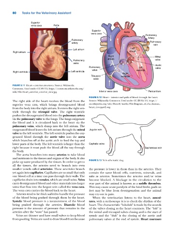
FIGURE 5.11 Heart – exterior structures. Source: Wikimedia
Commons. Used under CC BY 2.5, https://commons.wikimedia.org/
wiki/File:Heart_anterior_exterior_view.jpg. Inferior vena cava Pericardium
FIGURE 5.12 Heart – interior and path of blood through the heart.
The right side of the heart receives the blood from the Source: Wikimedia Commons. Used under CC BY‐SA 3.0, https://
superior vena cava, which brings deoxygenated blood en.wikipedia.org/wiki/Heart#/media/File:Diagram_of_the_human_
from the body into the right atrium. It enters the right ven- heart_(cropped).svg.
tricle through the tricuspid valve. The right ventricle
pushes the deoxygenated blood into the pulmonary artery
via the pulmonary valve to the lungs. The lungs oxygenate
the blood and it is circulated back to the heart via the
pulmonary veins, which dump into the left atrium. The
oxygenated blood leaves the left atrium through the mitral Jugular vein
valve to the left ventricle. The left ventricle pushes the oxy-
genated blood through the aortic valve into the aorta
which branches off at the aortic arch to feed the top and
lower parts of the body. The left ventricle is larger than the Cephalic veins
right because it must push the blood all the way through Saphenous vein
the body.
The aorta branches into many arteries to take blood
and nutrients to the tissues and organs of the body. It also
picks up waste produced by the tissues. In order to get to FIGURE 5.13 Vein schematic: dog.
all the tissues, the arteries need to branch into even
smaller vessels called arterioles. These arterioles branch the pressure is lower in them than in the arteries. They
yet again into capillaries. Capillaries are so small that only contain the same blood cells, nutrients, minerals, and
one blood cell at a time can pass through their walls. The salts as arteries. Sometimes the arteries and/or veins
capillaries drain into venules, which are small veins. Veins become blocked. A blockage in the circulation to the
carry deoxygenated blood and other materials into larger rear part of the animal is known as a saddle thrombus.
veins that flow into the largest vein called the vena cava. This may cause acute paralysis of the hind limbs, pads on
The vena cava carries the blood back to the heart. feet may be blue from deoxygenation and the animal
Arteries tend to be thick walled to handle the pressure may cry out in pain.
of the blood being pushed through them by the heart. When the veterinarian listens to the heart, auscul-
Systolic blood pressure is a measurement of the blood tates, with a stethoscope it is to check the rhythm of the
being pushed through the arteries. Diastolic blood heart. The characteristic “lub‐dub” is made by the sounds
pressure is the amount of pressure that remains in the of the valves closing as the heart contracts. The “lub” is
arteries after the “wave” has passed. the mitral and tricuspid valves closing and is the start of
Veins are thinner and have small valves to keep blood systole and the “dub” is the closing of the aortic and
from pooling. Veins are used to draw blood from because pulmonary valves at the end of systole. Heart murmurs