Page 272 - Essential Haematology
P. 272
258  /  Chapter 20  Non-Hodgkin lymphoma


                    (a)                                       (b)


                    (c)                                       (d)


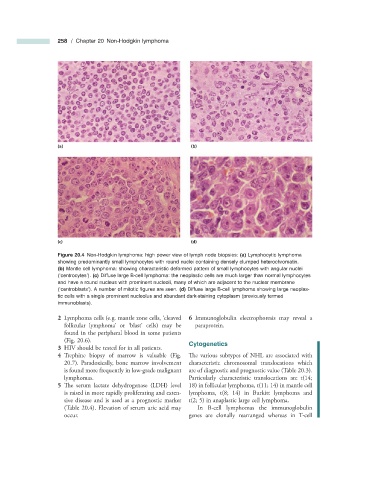
                              Figure 20.4   Non - Hodgkin lymphoma: high power view of lymph node biopsies:  (a)  Lymphocytic lymphoma
                    showing predominantly small lymphocytes with round nuclei containing densely clumped heterochromatin.
                      (b)  Mantle cell lymphoma: showing characteristic deformed pattern of small lymphocytes with angular nuclei
                    ( ‘ centrocytes ’ ).  (c)  Diffuse large B - cell lymphoma: the neoplastic cells are much larger than normal lymphocytes
                    and have a round nucleus with prominent nucleoli, many of which are adjacent to the nuclear membrane
                    ( ‘ centroblasts ’ ). A number of mitotic fi gures are seen.  (d)  Diffuse large B - cell lymphoma showing large neoplas-
                    tic cells with a single prominent nucleolus and abundant dark - staining cytoplasm (previously termed
                    immunoblasts).

                       2      Lymphoma cells (e.g. mantle zone cells,   cleaved      6      Immunoglobulin  electrophoresis  may  reveal  a
                                                     ‘
                      follicular  lymphoma ’   or   ‘ blast ’   cells)  may  be   paraprotein.
                      found in the peripheral blood in some patients
                      (Fig.  20.6 ).
                                                                  Cytogenetics

                       3   HIV should be tested for in all patients.


                       4      Trephine biopsy of marrow is valuable (Fig.    The various subtypes of NHL are associated with

                        20.7 ). Paradoxically, bone marrow involvement   characteristic chromosomal translocations which
                      is found more frequently in low - grade malignant   are of diagnostic and prognostic value (Table  20.3 ).
                      lymphomas.                              Particularly characteristic translocations are t(14;
                       5      The serum lactate dehydrogenase (LDH) level   18) in follicular lymphoma, t(11; 14) in mantle cell

                      is raised in more rapidly proliferating and exten-  lymphoma, t(8; 14) in Burkitt lymphoma and
                      sive disease and is used as a prognostic marker   t(2; 5) in anaplastic large cell lymphoma.
                      (Table  20.4 ). Elevation of serum uric acid may    In B - cell lymphomas the immunoglobulin
                      occur.                                  genes are clonally rearranged whereas in  T - cell

