
Page 274 - Essential Haematology
P. 274
260 / Chapter 20 Non-Hodgkin lymphoma
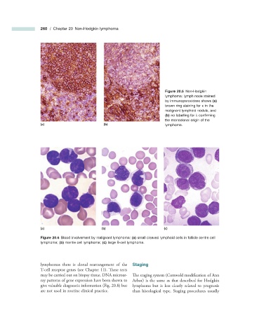
Figure 20.5 Non - Hodgkin
lymphoma: lymph node stained
by immunoperoxidase shows (a)
brown ring staining for κ in the
malignant lymphoid nodule, and
(b) no labelling for λ confi rming
the monoclonal origin of the
(a) (b) lymphoma.
(a) (b) (c)
Figure 20.6 Blood involvement by malignant lymphoma: (a) small cleaved lymphoid cells in follicle centre cell
lymphoma; (b) mantle cell lymphoma; (c) large B - cell lymphoma.
lymphomas there is clonal rearrangement of the Staging
T - cell receptor genes (see Chapter 11 ) . Th ese tests
may be carried out on biopsy tissue. DNA microar- The staging system (Cotswold modification of Ann
ray patterns of gene expression have been shown to Arbor) is the same as that described for Hodgkin
give valuable diagnostic information (Fig. 20.8 ) but lymphoma but is less clearly related to prognosis
are not used in routine clinical practice. than histological type. Staging procedures usually