
Page 278 - Essential Haematology
P. 278
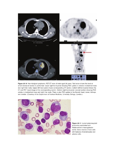
Figure 20.10   Non - Hodgkin lymphoma. PET/CT scan of male aged 26 years. Red arrow shows the level at
                    which transaxial section is performed. Upper right - hand panel showing FDG uptake in anterior mediastinal mass
                    and right hilar node. Upper left - hand panel shows corresponding CT section, bottom left - hand panel shows the
                    CT and PET fused image of the corresponding section. Bottom right - hand panel, coronal section showing FDG
                    uptake in mediastinal mass and right hilar node. There is also FDG uptake in the thyroid, heart, bowel, kidneys
                    and bladder.  (Courtesy of the Department of Nuclear Medicine, University College, London.)


                                                                                     Figure 20.11   Lymphoplasmacytoid
                                                                           lymphoma associated with
                                                                           Waldenstr ö m ’ s macroglobulin-
                                                                           aemia. Bone marrow shows cells
                                                                           with features of lymphocytes and
                                                                           plasma cells.