Page 279 - Essential Haematology
P. 279
Chapter 20  Non-Hodgkin lymphoma  /  265


                          Treatment                             germinal follicles. It is thought that lymphoid
                        No treatment is required for patients without symp-  hyperplasia initially occurs in response to antigen or
                      toms, but treatment should be started if there is  infl ammation and then cells acquire secondary

                      significant organomegaly, anaemia, thrombocyto-  genetic damage that leads to lymphoma. Th ey are
                      penia, neuropathy, amyloidosis or hyperviscosity.  classifi ed according the anatomical site at which
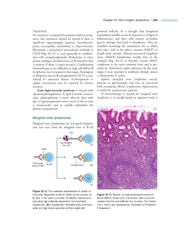
                      Rituximab, a humanized monoclonal antibody to  they arise, such as the spleen, mucosa (MALT) or
                      CD20 (Fig.  20.12 ), is used, generally in combina-  lymph node (nodal). Mucosa - associated lymphoid

                      tion with cyclophosphamide, fludarabine or other  tissue (MALT) lymphomas usually arise in the
                      purine analogue, bendamustine or bortezomib (but  stomach (Fig.  20.13 ) or thyroid. Gastric MALT
                      is omitted if there is hyperviscosity). Combination  lymphoma is the most common form and is pre-
                      chemotherapy as for follicular or large cell diff use B  ceded by  Helicobacter pylori  infection. In the early
                      lymphoma may be needed in late stages. Autologous  stages it may respond to antibiotic therapy aimed
                      or allogeneic stem cell transplantation (SCT) is con-  at eliminating  H. pylori .
                      sidered for advanced disease. Erythropoietin or    Splenic marginal zone lymphoma usually
                      regular transfusions may be required for chronic  presents as splenomegaly and may be associated
                      anaemia.                                  with circulating  ‘ villous ’  lymphocytes. Splenectomy
                            Acute hyperviscosity syndrome  is treated with  is useful for symptomatic patients.
                      repeated plasmapheresis. As IgM is mainly intravas-    If chemotherapy is needed for marginal zone
                      cular, plasmapheresis is more effective than with  lymphoma it is usually based on regimens used in

                      IgG or IgA paraproteins when much of the protein
                      is extravascular and so rapidly replenishes the
                      plasma compartment.


                          Marginal  z one  l ymphomas
                        Marginal zone lymphomas are low - grade lympho-
                      mas that arise from the marginal zone of B - cell

                                               FcR


                                         Neutrophil/
                                          NK cell           (a)
                            CD20


                       Rituximab
                               B cell          Complement   (b)
                      (Anti-CD20)


                                                            (c)


                                Figure 20.12   The potential mechanisms of action of


                      rituximab. Rituximab binds to CD20 on the surface of             Figure 20.13   Gastric mucosa - associated lymphoid
                      B cells. It can elicit a number of effector mechanisms   tissue (MALT) lymphoma: the tumour cells surround
                      including:  (a)  antibody dependent cell - mediated   reactive follicles and infi ltrate the mucosa. The follicle
                      cytotoxicity;  (b)  complement mediated lysis of tumour   has a  ‘ starry sky ’  appearance.  (Courtesy of Professor
                      cells; and  (c)  direct apoptosis of the target cell.     P. Isaacson.)

