
Page 385 - Essential Haematology
P. 385
Chapter 27 Thrombosis and antithrombotic therapy / 371
extent of the thrombus (Fig. 27.2 b). However, it is Chest X - ray This is often normal but may
a painful invasive technique, with a risk of contrast show evidence of pulmonary infarction or pleural
reaction and procedure - induced DVT. eff usion.
Plasma D - dimer concentration Th e concen- Ventilation perfusion (VQ) scintigraphy Th is
tration of these fibrin breakdown products is raised detects areas of the lung being ventilated but not
when there is a fresh thrombosis. It is a useful assay perfused.
when venous thrombosis is suspected and with the Computed tomography (CT) pulmonary
help of clinical probability shown by the Wells score angiography Fine slices of the lung are scanned
(Table 27.4 ). A negative result in emergency depart- by spiral CT so that filling defects in the pulmonary
ments can be used to exclude DVT. D - dimer eleva- arteries are visualized (Fig. 27.2 c).
tion in cancer, inflammation after surgery or trauma Magnetic resonance pulmonary angio-
limits its usefulness. graphy Gadolinium - enhanced MRI is a relatively
Magnetic resonance imaging (MRI) Th is new, expensive but accurate technique.
may also be used but is expensive. Impedance Pulmonary angiography This is the tradi-
plethysmography is less sensitive and accurate and tional reference method but is invasive with com-
is falling out of use. plications, albeit uncommon, such as arrhythmia or
contrast reaction.
Electrocardiogram This is performed to
Pulmonary e mbolus
‘
determine whether there is right heart strain ’ which
Clinical suspicion This is particularly suspected occurs only in relatively severe cases.
in patients with chest symptoms, especially if there
are signs, or previous history of DVT, immobiliza-
tion for more than 2 days or recent ( < 4 weeks) Anticoagulant d rugs
surgery, haemoptysis or cancer. Recurrent PE may
Anticoagulant drugs are used widely in the treat-
lead to pulmonary hypertension.
ment of venous thromboembolic disease. Th eir
value in the treatment of arterial thrombosis is less
well established.
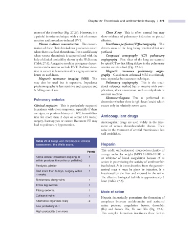
Table 27.4 Deep vein thrombosis: clinical
assessment: the Wells score. Heparin
This acidic unfractionated mucopolysaccharide of
Points
average molecular weight (MW) 15 000 – 18 000 is
Active cancer (treatment ongoing or 1 an inhibitor of blood coagulation because of its
within previous 6 months or palliative)
action in potentiating the activity of antithrombin
Paralysis, plaster 1 (see below). As it is not absorbed from the gastroin-
testinal tract it must be given by injection. It is
Bed more than 3 days, surgery within 1
4 weeks inactivated by the liver and excreted in the urine.
Th e effective biological half - life is approximately 1
Tenderness along veins 1
hour (Table 27.5 ).
Entire leg swollen 1
Pitting oedema 1
Mode of a ction
Collateral veins 1
Heparin dramatically potentiates the formation of
Alternative diganosis likely – 2 complexes between antithrombin and activated
Low probability 0 – 1 serine protease coagulation factors, thrombin
(IIa) and factors IXa, Xa and XIa (Fig. 27.6 ).
High probability 2 or more
This complex formation inactivates these factors