
Page 48 - Essential Haematology
P. 48
34 / Chapter 3 Hypochromic anaemias
Iron deficiency is the most common cause of Body i ron d istribution and t ransport
anaemia in every country of the world. It is
The transport and storage of iron is largely mediated
the most important cause of a microcytic
by three proteins: transferrin, transferrin receptor 1
hypochromic anaemia, in which the two red cell
(TfR1) and ferritin.
indices, mean corpuscular volume (MCV) and
Transferrin can contain up to two atoms of iron.
mean corpuscular haemoglobin (MCH), are
It delivers iron to tissues that have transferrin recep-
reduced and the blood film shows small (micro-
tors, especially erythroblasts in the bone marrow
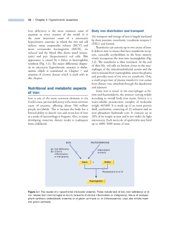
cytic) and pale (hypochromic) red cells. Th is
which incorporate the iron into haemoglobin (Fig.
appearance is caused by a defect in haemoglobin
3.2 ). The transferrin is then reutilized. At the end
synthesis (Fig. 3.1 ). The major diff erential diagno-
of their life, red cells are broken down in the mac-
sis in microcytic hypochromic anaemia is thalas-
rophages of the reticuloendothelial system and the
saemia which is considered in Chapter 7 and
iron is released from haemoglobin, enters the plasma
anaemia of chronic disease which is dealt with in
and provides most of the iron on transferrin. Only
this chapter.
a small proportion of plasma transferrin iron comes
from dietary iron, absorbed through the duodenum
Nutritional and m etabolic a spects and jejunum.
Some iron is stored in the macrophages as fer-
of i ron
ritin and haemosiderin, the amount varying widely
Iron is one of the most common elements in the according to overall body iron status. Ferritin is a
’
Earth s crust, yet iron deficiency is the most common water - soluble protein – iron complex of molecular
cause of anaemia, affecting about 500 million weight 465 000. It is made up of an outer protein
people worldwide. This is because the body has a shell, apoferritin, consisting of 22 subunits and an
limited ability to absorb iron and excess loss of iron iron – phosphate – hydroxide core. It contains up to
as a result of haemorrhage is frequent. Also, in many 20% of its weight as iron and is not visible by light
developing countries, dietary intake is inadequate microscopy. Each molecule of apoferritin may bind
from childhood. up to 4000 – 5000 atoms of iron.
IRON PROTOPORPHYRIN
(a) Iron deficiency Sideroblastic
(b) Chronic anaemia
inflammation
or malignancy
Haem + Globin
Thalassaemia (α or β)
Haemoglobin
Figure 3.1 The causes of a hypochromic microcytic anaemia. These include lack of iron (iron defi ciency) or of
iron release from macrophages to serum (anaemia of chronic infl ammation or malignancy), failure of protopor-
phyrin synthesis (sideroblastic anaemia) or of globin synthesis ( α - or β - thalassaemia). Lead also inhibits haem
and globin synthesis.