
Page 184 - parasitology for medical and clinical laboratoryprofessionals
P. 184
164 CHAPTER 7
Epidemiology of Cysticercosis
In the sixteenth and seventeenth centuries, structures
resembling cysticerci were documented by several
scientists, but none of the documentation of these obser-
vations initially suggested that parasites were responsible
for the formation of these cysts. It was not until the late Source: Centers for Disease Control and Prevention (CDC)
eighteenth century when Johann Goeze demonstrated
that certain cysts were the larval stages of tapeworms.
A major medical complication associated with infection
of T. solium is cysticercosis, where the infected individ-
ual becomes the intermediate host and harbors the larvae
in tissues throughout the body. Therefore, T. solium may
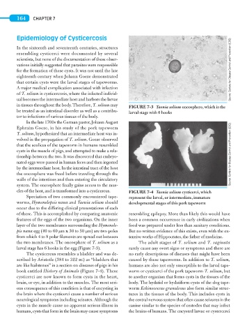
FIGURE 7-3 Taenia solium oncosphere, which is the
be treated as an intestinal disorder as well as a contribu- larval stage with 6 hooks
tor to infections of various tissues of the body.
In the late 1700s the German pastor, Johann August
Ephraim Goeze, in his study of the pork tapeworm
T. solium, hypothesized that an intermediate host was in-
volved in the propagation of T. solium. Goeze observed
that the scolices of the tapeworm in humans resembled
cysts in the muscle of pigs, and attempted to make a rela-
tionship between the two. It was discovered that embryo- Source: Centers for Disease Control and Prevention (CDC)
nated eggs were passed in human feces and then ingested
by the intermediate host. In the intestinal tract of the host
the oncosphere was freed before traveling through the
walls of the intestines and then entering the circulatory
system. The oncosphere finally gains access to the mus-
cles of the host, and is transformed into a cysticercus. FIGURE 7-4 Taenia solium cysticerci, which
Speciation of two commonly encountered tape- represent the larval, or intermediate, immature
worms, Hymenolepsis nana and Taenia solium should developmental stages of this pork tapeworm
occur due to the differing clinical presentations of each
of these. This is accomplished by comparing anatomic resembling epilepsy. More than likely this would have
features of the eggs of the two organisms. On the inner been a common occurrence in early civilizations when
layer of the two membranes surrounding the Hymenole- food was prepared under less than sanitary conditions.
pis nana egg (40 to 60 μm x 30 to 50 μm) are two poles But no written evidence of this exists, even with the ex-
from which 4 to 8 polar filaments are spread out between tensive works of Hippocrates, the father of medicine.
the two membranes. The oncosphere of T. solium as a The adult stages of T. solium and T. saginata
larval stage has 6 hooks in the egg (Figure 7-3). rarely cause any overt signs or symptoms and there are
The cysticercus resembles a bladder and was de- no early descriptions of diseases that might have been
scribed by Aristotle (384 to 322 bc) as “bladders that caused by these tapeworms. In addition to T. solium,
are like hailstones” in a section on diseases of pigs in his humans are also not only susceptible to the larval tape-
book entitled History of Animals (Figure 7-4). These worm or cysticerci of the pork tapeworm T. solium, but
cysticerci are now known to form cysts in the heart, to another organism that forms cysts in the tissues of the
brain, or eye, in addition to the muscles. The most seri- body. The hydatid or hydatiform cysts of the dog tape-
ous consequence of this condition is that of encysting in worm Echinococcus granulosus also form similar struc-
the brain where the cysticerci cause a number of serious tures in the tissues of the body. This includes cysts in
neurological symptoms including seizures. Although the the central nervous system that often cause seizures in the
cysts in the muscle cause no apparent serious illness in canine similar to the species of cestodes that may infect
humans, cysts that form in the brain may cause symptoms the brains of humans. The encysted larvae or cystercerci