
Page 185 - parasitology for medical and clinical laboratoryprofessionals
P. 185
Intestinal Cestodes 165
of T. solium in the flesh of pigs were known in ancient new proglottids form in the neck region. The segments
history as “measly pork” and appeared to be well known nearest the neck are immature and in this region the sex
to the ancient Greek physicians, but the condition was organs not fully developed, whereas those closer to the
never described as a disease of humans. posterior end are mature and are fully capable of repro-
Evidence exists from early history in the Bible of duction. The terminal segments are gravid (pregnant)
the Jews regarding the dangers associated with eating and the egg-filled uterus of each segment is the most
certain foods. Certain dietary restrictions abound in the prominent feature.
Jewish faith as well as for the Muslims as two religious
groups that also observe similar proscriptions against
eating pork and who practice the ritualistic preparation DIPHYLLOBOTHRIUM LATUM
of foods. Indirect evidence from different cultures indi-
cates that people were aware of the possible dangers in- Humans may also harbor the adults of Diphyllobothrium
herent in eating the flesh of pigs because infections with latum, the broad tapeworm that is also called the fish
cysticerci are rarely found in Jews and Muslims. tapeworm, which lives in the intestine. Eggs are passed
By the nineteenth century, animal experiments dem- in the feces and the first larval stage, the coracidium,
onstrated without a doubt that cysticercosis was caused by develops within the egg and when eaten by a copepod
the ingestion of T. solium eggs. These observations led to (a minute crustacean found in fresh water), it proceeds
public health measures, which had a significant impact on to develop into the second larval stage called the
the control of tapeworm infections in humans by restricting procercoid. When an infected copepod is eaten by a
the amount infected meat on the market for human con- fish, the procercoid develops into the third larval stage,
sumption. Because meat was required to be inspected be- the plerocercoid, and when a human eats an infected
fore sale as long as 200 years ago, those who ate meat from fish, the plerocercoid develops into an adult tapeworm
the butcher shops may have been relatively safe. But many in the gut (Figure 7-6). This particular tapeworm
poor farmers raised their own meat and almost certainly was well known in ancient history and is mentioned,
fed their animals contaminated food, which their families sometimes indirectly, in the major classical medical
ate and was a practice that propagated cysticercosis. writings including the Ebers papyrus, the Corpus
Hippocratorum, and the works of Celsus and Avicenna
Identification of Cestodes (Cox, 2002). However, there are no accurate early
clinical records because there are few overt signs of the
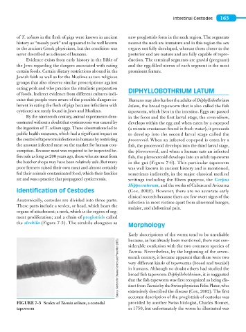
Anatomically, cestodes are divided into three parts.
infection in most victims apart from abnormal hunger,
These parts include a scolex, or head, which bears the
malaise, and abdominal pain.
organs of attachment; a neck, which is the region of seg-
ment proliferation; and a chain of proglottids called
the strobila (Figure 7-5). The strobila elongates as
Morphology
Early descriptions of the worm tend to be unreliable
because, as has already been mentioned, there was con-
Source: Centers for Disease Control and Prevention (CDC) Taenia. Nevertheless, by the beginning of the seven-
siderable confusion with the two common species of
teenth century, it became apparent that there were two
very different kinds of tapeworms (broad and taeniid)
in humans. Although no doubt others had studied the
broad fish tapeworm Diphyllobothrium, it is suggested
that the fish tapeworm was first recognized as being dis-
extensively described the disease (Cox, 2002). The first
accurate description of the proglottids of cestodes was
provided by another Swiss biologist, Charles Bonnet,
FIGURE 7-5 Scolex of Taenia solium, a cestodal tinct from Taenia by the Swiss physician Felix Plater, who
tapeworm in 1750, but unfortunately the worm he illustrated was