
Page 19 - Live-cellanalysis handbook
P. 19
Kinetic Proliferation Assays
®
IncuCyte Proliferation Assays at a glance
A variety of strategies for kinetic measurement of proliferation are to identify in phase contrast images, IncuCyte NucLight live-
possible using the IncuCyte Live-Cell Analysis System. Selection cell analysis reagents can be used to fluorescently label nuclei.
of label-free or fluorescent assays depends on the specific Fluorescent IncuCyte images can then be acquired over time and
scientific question being asked and cell models studied. Continuous analyzed to generate nuclear counts and derive doubling times in
live-cell assays for both adherent and non-adherent cells are either mono- or co-cultures.
possible, as cells stay stationary inside a standard tissue culture
incubator while IncuCyte® optics move. There is no sample or stage Additionally, IncuCyte Proliferation Assays can be multiplexed
movement that can cause non-adherent cells to migrate to the with IncuCyte Live-Cell Analysis fluorescence reagents for cell
edges of microplate wells and negatively impact data accuracy. health assessments, including apoptosis (IncuCyte® Caspase
3/7, Annexin V), cytotoxicity (IncuCyte® Cytotox), or viability
IncuCyte® Live-Cell Imaging and Analyis enables non-invasive, (IncuCyte® NucLight Lentivirus). Cell boundaries can be identified
label-free measurements of cell growth based on area (confluence) using the IncuCyte® Cell-by-Cell Analysis software tools, and
or cell number (count) metrics, both of which are generated via simultaneous assessment of cell death or viability achieved by
segmentation (masking) of high quality phase images. To resolve measuring the fluorescence intensity originating from within the
the challenge of quantifying low contrast cells that can be difficult individual cell boundary.
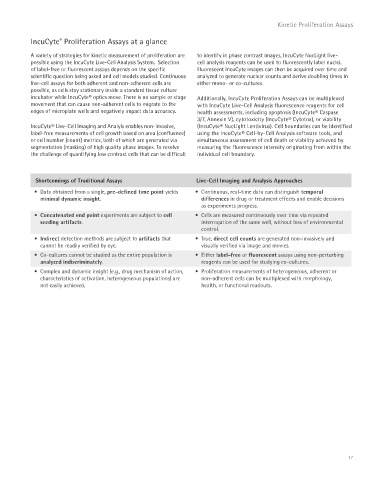
Shortcomings of Traditional Assays Live-Cell Imaging and Analysis Approaches
• Data obtained from a single, pre-defined time point yields • Continuous, real-time data can distinguish temporal
minimal dynamic insight. differences in drug or treatment effects and enable decisions
as experiments progress.
• Concatenated end point experiments are subject to cell • Cells are measured continuously over time via repeated
seeding artifacts. interrogation of the same well, without loss of environmental
control.
• Indirect detection methods are subject to artifacts that • True, direct cell counts are generated non-invasively and
cannot be readily verified by eye. visually verified via image and movies.
• Co-cultures cannot be studied as the entire population is • Either label-free or fluorescent assays using non-perturbing
analyzed indiscriminately. reagents can be used for studying co-cultures.
• Complex and dynamic insight (e.g., drug mechanism of action, • Proliferation measurements of heterogeneous, adherent or
characteristics of activation, heterogeneous populations) are non-adherent cells can be multiplexed with morphology,
not easily achieved. health, or functional readouts.
17