
Page 22 - Live-cellanalysis handbook
P. 22
Live-Cell Analysis Handbook — Third Edition
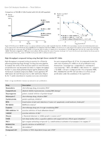
Comparison of SK-BR-3 Cells Treated with 555.56 nM Lapatinib
B Nuclear count/mm 2
A Nuclear 3
count/mm 2 (AUC x 10 )
200
1500
SK-BR-3
+ CCD-1068Sk
150
1000
SK-BR-3 + HMF 100
500 SK-BR-3 without
Stromal Cells 50 SK-BR-3 + CCD-1068Sk (IC = 1.137 μM)
50
SK-BR-3 + HMF (IC = 0.581 μM)
50
SK-BR-3 without Stromal Cells (IC = 0.015 μM)
0 0 50
0 50 100 150 200 -8 -7 -6 -5
Time (h) Log [Lapatinib] M
10
Figure 4. Proliferation of SK-BR-3 cells in co-culture and monoculture under lapatinib treatment. SK-BR-3 cells expressing a nuclear restricted red protein were
grown with normal skin fibroblasts (CCD-1068Sk), human mammary fibroblasts (HMF), or in monoculture, and treated with varying concentrations of lapatinib for
8 days. (A) Nuclear counts per mm of SK-BR-3 cells grown with or without stromal cells in the presence of 556 nM lapatinib illustrate the rescue effect of CCD-
2
2
1068Sk fibroblasts compared to HMFs and mono-culture. (B) Area under the curve of nuclear counts per mm over time for each concentration (n=4) was used to
calculate and compare IC values of SK-BR-3 cells grown with or without stromal cells.
50
High-throughput compound testing using NucLight Green labeled HT-1080s
High-throughput compound testing is essential for efficiently for each compound (Figure 6). Of the 16 compounds tested, the
advancing promising drugs through the drug discovery pipeline. rank order of potency for inhibition of cell proliferation was:
To examine the ability of the IncuCyte system to meet this need, doxorubicin = staurosporine = camptothecin > mitomycin C
cell proliferation was measured over time in a higher-throughput >cycloheximide = RITA > PD-98059 > FAK inhibitor 14 = cisplatin
format. To assess many pharmacological agents simultaneously, > 10-DEBC = Chrysin = Compound 401. The compounds TAME,
16 literature-standard compounds (Table 1) were applied to HT- PAC1, KU0063794 and FPA-124 had little or no effect on cell
1080 tumor-derived fibrosarcoma in a 384-well format (Figure proliferation under the conditions of the experiment.
5). An 11-point concentration response curve was constructed
Table 1. Drugs identified in literature as relevant to cell proliferation.
Drug Description
Doxorubicin chemotherapy drug, intercalates DNA 3
Camptothesin alkaloid inhibits topoisomerase, causing DNA damage 3
Staurosporine potent alkaloid inhibitor of protein kinase 4
Mitomycin C chemotherapy drug, alkylates DNA 5
Cycloheximide protein synthesis, inhibitor 6
RITA (reactivation of p53 and induction of tumor cell apoptosis) a small molecule, binds p53 7
PD-98059 MAPK1/2 inhibitor 8
Cisplatin chemotherapy drug acts through crosslinking DNA 9
FAK-inhibitor 14 selective inhibitor of focal adhension kinase 10
10DEBC selective inhibitor of Akt 11
Chrysin a flavonoid observed to inhibit growth in cancer cells 12
TAME tert-Amyl methyl ether; a gasoline additive with suspected toxic effects upon inhalation 13
PAC1 (pro-caspase activating compound-1), a small-molecule activator of procaspase-3 to caspase-3 14
KU0063794 specific inhibitor of mTORC1/2 15
FPA-124 Akt inhibitor 16
Compound 401 inhibitor of DNA-dependent kinase and mTOR 17
20