
Page 21 - Live-cellanalysis handbook
P. 21
Kinetic Proliferation Assays
If a label-free technique does not suffice, nuclear counts can be with minimal toxicity. These reagents are ideal for generating
performed using IncuCyte NucLight Reagents. These reagents are stable cell populations or clones using puromycin or bleomycin
available in either dye (IncuCyte® NucLight Rapid) or lentiviral selection. Fluorescent techniques have particular value when
(IncuCyte® NucLight Lentivirus) formats and are non-perturbing morphology of cells are flat and/or thin, and therefore IncuCyte
to cell health and morphology. IncuCyte NucLight Rapid is a NucLight lentivirus reagent can be utilized not only to determine
cell permeable DNA stain that specifically stains nuclei in cells nuclear counts in living cells as described in Figure 3, but also
using a mix-and-read protocol. IncuCyte NucLight Lentivirus in combination with label-free cell identification methods to
Reagents are compatible with convenient transduction protocols measure viability (data not shown). Loss of viability is indicated
and provide homogenous expression of a nuclear-restricted by a loss in fluorescence, as fluorescent protein passes out of the
fluorescent protein in your choice of primary, immortalized, nucleus as nuclear membrane integrity is lost.
dividing, or non-dividing cells without altering cell function and
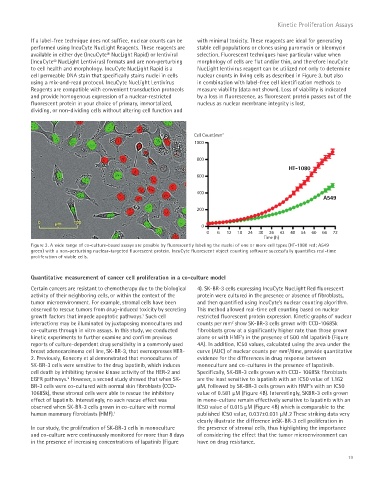
Cell Count/mm 2
1000
800
HT-1080
600
400
A549
200
0
0 6 12 18 24 30 36 42 48 54 60 66 72
Time (h)
Figure 3. A wide range of co-culture-based assays are possible by fluorescently labeling the nuclei of one or more cell types (HT-1080 red; A549
green) with a non-perturbing nuclear-targeted fluorescent protein. IncuCyte fluorescent object counting software successfully quantifies real-time
proliferation of viable cells.
Quantitative measurement of cancer cell proliferation in a co-culture model
Certain cancers are resistant to chemotherapy due to the biological 4). SK-BR-3 cells expressing IncuCyte NucLight Red fluorescent
activity of their neighboring cells, or within the context of the protein were cultured in the presence or absence of fibroblasts,
tumor microenvironment. For example, stromal cells have been and then quantified using IncuCyte’s nuclear counting algorithm.
observed to rescue tumors from drug-induced toxicity by secreting This method allowed real-time cell counting based on nuclear
growth factors that impede apoptotic pathways. Such cell restricted fluorescent protein expression. Kinetic graphs of nuclear
1
interactions may be illuminated by juxtaposing monocultures and counts per mm show SK-BR-3 cells grown with CCD-1068Sk
2
co-cultures through in vitro assays. In this study, we conducted fibroblasts grow at a significantly higher rate than those grown
kinetic experiments to further examine and confirm previous alone or with HMF’s in the presence of 500 nM lapatinib (Figure
reports of culture-dependent drug sensitivity in a commonly used 4A). In addition, IC50 values, calculated using the area under the
2
breast adenocarcinoma cell line, SK-BR-3, that overexpresses HER- curve (AUC) of nuclear counts per mm /time, provide quantitative
2. Previously, Konecny et al demonstrated that monocultures of evidence for the differences in drug response between
SK-BR-3 cells were sensitive to the drug lapatinib, which induces monoculture and co-cultures in the presence of lapatinib.
cell death by inhibiting tyrosine kinase activity of the HER-2 and Specifically, SK-BR-3 cells grown with CCD- 1068Sk fibroblasts
EGFR pathways. However, a second study showed that when SK- are the least sensitive to lapatinib with an IC50 value of 1.162
2
BR-3 cells were co-cultured with normal skin fibroblasts (CCD- μM, followed by SK-BR-3 cells grown with HMF’s with an IC50
1068Sk), these stromal cells were able to rescue the inhibitory value of 0.581 μM (Figure 4B). Interestingly, SKBR-3 cells grown
effect of lapatinib. Interestingly, no such rescue effect was in mono-culture remain effectively sensitive to lapatinib with an
observed when SK-BR-3 cells grown in co-culture with normal IC50 value of 0.015 μM (Figure 4B) which is comparable to the
human mammary fibroblasts (HMF). published IC50 value, 0.037±0.031 μM.2 These striking data very
1
clearly illustrate the difference inSK-BR-3 cell proliferation in
In our study, the proliferation of SK-BR-3 cells in monoculture the presence of stromal cells, thus highlighting the importance
and co-culture were continuously monitored for more than 8 days of considering the effect that the tumor microenvironment can
in the presence of increasing concentrations of lapatinib (Figure have on drug resistance.
19