
Page 32 - Rapid Review of ECG Interpretation in Small Animal Practice, 2nd Edition
P. 32
Evaluation of the Electrocardiogram
VetBooks.ir I aVR
aVL
II
aVF
III
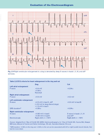
Fig. 2.9 Right ventricular enlargement in a dog is denoted by deep S waves in leads I, II, III, and aVF
(arrows).
Table 2.2 ECG criteria for heart enlargement in the dog and cat
Dog Cat
Left atrial enlargement
P wave >0.4 mV >0.04 s
>0.04 s
Notched
Right atrial enlargement
P wave >0.4 mV >0.2 mV
Left ventricular enlargement
R wave >2.5 mV in lead II, aVF >0.9 mV in lead II
(>3.0 mV in large breed dogs)
>1.5 mV in lead I
QRS duration* >0.06 s >0.04 s
Right ventricular enlargement
S wave >0.05 mV in lead I S wave in leads I, II, III
>0.35 mV in lead II and aVF
Electrical axis Right shift (>+100°) Right shift (>+160°)
Source: Adapted from Tilley LP, Smith WK (2008). Electrocardiography. In: Tilley LP, Smith WK, Oyama MA, Sleeper
MM (eds). Manual of Canine and Feline Cardiology, 4th edn. Saunders Elsevier, St Louis.
*QRS duration >0.08 s in the dog and >0.06 s in the cat can be associated with left or right bundle branch blocks. See
text for more detail.
19