
Page 327 - Zoo Animal Learning and Training
P. 327
Chapter 16 Diagnostic Imaging and Endoscopy 311
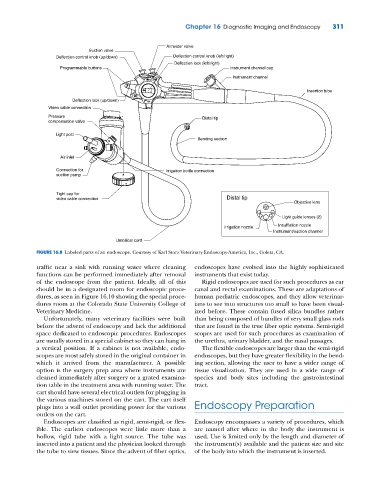
Air/water valve
Suction valve
Deflection control knob (up/down) Deflection control knob (left/right)
Deflection lock (left/right)
Programmable buttons Instrument channel cap
Instrument channel
Insertion tube
Deflection lock (up/down)
Video cable connection
Pressure Distal tip
compensation valve
Light post
Bending section
Air inlet
Connection for Irrigation bottle connection
suction pump
Tight cap for
video cable connection Distal tip
Objective lens
Light guide lenses (2)
Irrigation nozzle Insufflation nozzle
Instrument/suction channel
Umbilical cord
FIGURE 16.8 Labeled parts of an endoscope. Courtesy of Karl Storz Veterinary Endoscopy‐America, Inc., Goleta, CA.
traffic near a sink with running water where cleaning endoscopes have evolved into the highly sophisticated
functions can be performed immediately after removal instruments that exist today.
of the endoscope from the patient. Ideally, all of this Rigid endoscopes are used for such procedures as ear
should be in a designated room for endoscopic proce- canal and rectal examinations. These are adaptations of
dures, as seen in Figure 16.10 showing the special proce- human pediatric endoscopes, and they allow veterinar-
dures room at the Colorado State University College of ians to see into structures too small to have been visual-
Veterinary Medicine. ized before. These contain fused silica bundles rather
Unfortunately, many veterinary facilities were built than being composed of bundles of very small glass rods
before the advent of endoscopy and lack the additional that are found in the true fiber optic systems. Semi‐rigid
space dedicated to endoscopic procedures. Endoscopes scopes are used for such procedures as examination of
are usually stored in a special cabinet so they can hang in the urethra, urinary bladder, and the nasal passages.
a vertical position. If a cabinet is not available, endo- The flexible endoscopes are larger than the semi‐rigid
scopes are most safely stored in the original container in endoscopes, but they have greater flexibility in the bend-
which it arrived from the manufacturer. A possible ing section, allowing the user to have a wider range of
option is the surgery prep area where instruments are tissue visualization. They are used in a wide range of
cleaned immediately after surgery or a grated examina- species and body sites including the gastrointestinal
tion table in the treatment area with running water. The tract.
cart should have several electrical outlets for plugging in
the various machines stored on the cart. The cart itself
plugs into a wall outlet providing power for the various Endoscopy Preparation
outlets on the cart.
Endoscopes are classified as rigid, semi‐rigid, or flex- Endoscopy encompasses a variety of procedures, which
ible. The earliest endoscopes were little more than a are named after where in the body the instrument is
hollow, rigid tube with a light source. The tube was used. Use is limited only by the length and diameter of
inserted into a patient and the physician looked through the instrument(s) available and the patient size and site
the tube to view tissues. Since the advent of fiber optics, of the body into which the instrument is inserted.