
Page 329 - Zoo Animal Learning and Training
P. 329
Chapter 16 Diagnostic Imaging and Endoscopy 313
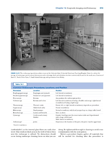
FIGURE 16.10 The endoscopy/special procedures room at the Colorado State University Veterinary Teaching Hospital. Note the cabinet for
storage of endoscopes and accessory instruments and a videotape deck and television monitor conveniently located in the work area. Courtesy of
David C. Twedt. From Tams, T. Small Animal Endoscopy, with permission from Mosby.
Table 16.1
Common Endoscopic Procedures, Locations, and Position
Procedure Location Position
Esophagogastroscopy Esophagus and stomach Left lateral recumbency
Duodenojejunoscopy Duodenum and jejunum Left lateral recumbency
Proctoscopy Rectum Any position the endoscopist prefers
Colonoscopy Rectum and colon Left lateral recumbency if using a flexible endoscope; right lateral
recumbency if using a rigid scope
Thoracoscopy Thoracic cavity Dorsal, left, or right lateral recumbency; depends on procedure
Laparoscopy Abdominal cavity Dorsal recumbency
Rhinoscopy Nasal passages Sternal recumbency with head propped up on a large rolled towel
Bronchoscopy Larynx and bronchi Same as above
Cytoscopy Urethra and urinary Female: hind legs over the examination table and hips elevated
bladder with a large rolled towel
Male: lateral recumbency
Arthroscopy a Joints Depends on the location of the joint; the joint must be uppermost
a Requires surgical prep.
(unthinkable!) as the internal glass fibers are easily shat- doing. Be vigilant and thorough in cleaning to avoid cross‐
tered. This results in black areas in the field of vision when contamination with the next patient.
next the endoscope is utilized. No distractions should Before a procedure begins, gather all materials that
occur during endoscope cleaning; focus on what you are will be needed for cleaning after the procedure is