Page 238 - Essential Haematology
P. 238
224  /  Chapter 17  Acute lymphoblastic leukaemia


                     Acute lymphoblastic leukaemia (ALL) is caused by

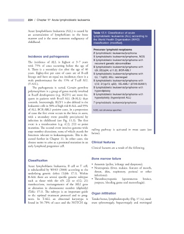
                                                                   Table 17.1   Classifi cation of acute
                    an accumulation of lymphoblasts in the bone
                                                               lymphoblastic leukaemia (ALL) according to
                    marrow and is the most common malignancy of
                                                               the World Health Organization (WHO)
                    childhood.                                 classifi cation (modifi ed).
                                                                     Precursor lymphoid neoplasms
                                                                     B lymphoblastic leukaemia/lymphoma
                        Incidence and  p athogenesis               B lymphoblastic leukaemia/lymphoma, NOS
                                                                   B lymphoblastic leukaemia/lymphoma with

                     The incidence of ALL is highest at 3 – 7 years
                                                                recurrent genetic abnormalities
                    with 75% of cases occurring before the age of
                                                                   B lymphoblastic leukaemia/lymphoma with

                    6. There is a secondary rise after the age of 40   t(9; 22)(q34; q11.2);  BCR - ABL1
                    years.  Eighty - five per cent of cases are of B - cell       B lymphoblastic leukaemia/lymphoma with

                    lineage and have an equal sex incidence; there is a   t(v; 11q23);  MLL  rearranged
                    male predominance for the 15% of  T - cell ALL       B lymphoblastic leukaemia/lymphoma with
                    (T - ALL).                                  t(12; 21)(p13; q22);  TEL - AML1  ( ETV6 - RUNX1 )
                       The pathogenesis is varied. Certain germline       B lymphoblastic leukaemia/lymphoma with

                    polymorphism in a group of genes mainly involved   hyperdiploidy
                    in B - cell development (e.g. IKZF1) are more fre-      B lymphoblastic leukaemia/lymphoma with
                    quent in patients with B - cell ALL (B - ALL) than   hypodiploidy (hypodiploid ALL)
                    controls. Interestingly, IKZF1 is also deleted in the         T lymphoblastic leukaemia/lymphoma
                    leukaemic cells in 30% of high risk B - ALL and 95%
                    of ALL BCR - ABL1 positive cases. In a proportion         NOS, not otherwise specifi ed.
                    of cases the first event occurs in the fetus  in utero ,

                    with a secondary event possibly precipitated by
                    infection in childhood (see Fig.  11.3   ). Th e  fi rst
                    event is a translocation (e.g. t(12; 21)) or point

                    mutation. The second event involves genome - wide
                                                              nalling pathway is activated in most cases (see
                    copy number alterations, some of which encode for
                                                              below).
                    functions relevant to leukaemogenesis. This is dis-


                    cussed further in Chapter  11 . In other cases, the
                    disease seems to arise as a postnatal mutation in an       Clinical  f eatures
                    early lymphoid progenitor cell.
                                                                Clinical features are a result of the following.
                                                                  Bone  m arrow  f ailure
                        Classifi cation
                                                                   •       Anaemia (pallor, lethargy and dyspnoea);
                      Acute lymphoblastic leukaemia, B cell or  T cell,
                                                                 •       Neutropenia  (fever,  malaise,  features  of  mouth,
                    is subclassified by WHO (2008) according to the

                                                               throat, skin, respiratory, perianal or other
                    underlying genetic defect (Table  17.1 ).  Within
                                                               infections);
                    B - ALL there are several specific genetic subtypes

                                                                 •       Th rombocytopenia  (spontaneous  bruises,
                    such as those with the t(9; 22) or t(12; 21)
                                                               purpura, bleeding gums and menorrhagia).
                    translocations, rearrangements of the  MLL   gene
                    or alteration in chromosome number (diploidy)
                    (Table   17.1 ).  The subtype is an important guide       Organ  i nfi ltration

                    to the optimal treatment protocol and to prog-
                    nosis. In  T - ALL an abnormal karyotype is     Tender bones, lymphadenopathy (Fig.  17.1 a), mod-
                    found in 50 – 70% of cases and the NOTCH sig-  erate splenomegaly, hepatomegaly and meningeal

