Page 250 - Essential Haematology
P. 250

236  /  Chapter 18  Chronic lymphoid leukaemias


                              Figure 18.1   Chronic lymphocytic leukaemia: bilateral             Figure 18.2   Chronic lymphocytic leukaemia: herpes

                    cervical lymphadenopathy in a 67 - year - old woman.   zoster infection in a 68 - year - old female.
                                                         9
                    Haemoglobin 12.5   g/dL; white blood count 150    ×   10  /L

                                    9
                                                    9


                    (lymphocytes 146    ×   10  /L); platelets 120    ×   10  /L.


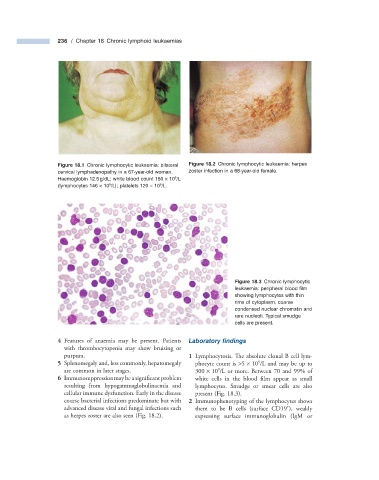
                                                                                      Figure 18.3   Chronic lymphocytic


                                                                            leukaemia: peripheral blood fi lm
                                                                            showing lymphocytes with thin
                                                                            rims of cytoplasm, coarse
                                                                            condensed nuclear chromatin and
                                                                            rare nucleoli. Typical smudge
                                                                            cells are present.


                       4      Features of anaemia may be present. Patients         Laboratory  fi ndings
                      with thrombocytopenia may show bruising or
                      purpura.                                     1      Lymphocytosis. The absolute clonal B cell lym-

                                                                                    9
                       5      Splenomegaly and, less commonly, hepatomegaly   phocyte count is  > 5    ×    10  /L and may be up to
                                                                       9
                      are common in later stages.               300    ×    10  /L or more. Between 70 and 99% of
                       6      Immunosuppression may be a signifi cant problem   white cells in the blood film appear as small

                      resulting from hypogammaglobulinaemia and   lymphocytes. Smudge or smear cells are also
                      cellular immune dysfunction. Early in the disease   present (Fig.  18.3 ).
                      course bacterial infections predominate but with      2      Immunophenotyping of the lymphocytes shows
                                                                                               +
                      advanced disease viral and fungal infections such   them to be B cells (surface CD19  ),  weakly
                      as herpes zoster are also seen (Fig.  18.2 ).          expressing surface immunoglobulin (IgM or
   245   246   247   248   249   250   251   252   253   254   255