
Page 291 - Essential Haematology
P. 291
Chapter 21 Multiple myeloma and related disorders / 277
immune paresis . The urine contains free light per cent have no bone lesions. In addition,
chains, Bence - Jones protein , in two - thirds of pathological fractures or vertebral collapse (Fig.
cases. Rare cases of myeloma are non - secretory 21.3 b) are common. The osteolytic lesions are
and therefore not associated with a paraprotein caused by osteoclast activation resulting from
or Bence - Jones proteinuria although some will high serum levels of RANKL (receptor activator
still show a disturbed free light chain ratio in of nuclear factor - κ B (NF - κ B) ligand), pro-
the serum. duced by plasma cells and bone marrow stroma,
4 There is usually a normochromic, normocytic which binds to activatory RANK receptors on
or macrocytic anaemia. Rouleaux formation is the osteoclast surface.
marked in most cases (Fig. 21.5 ). Neutropenia 8 Serum calcium elevation occurs in 45% of
and thrombocytopenia occur in advanced patients. Typically, the serum alkaline phos-
disease. Abnormal plasma cells appear in the
blood film in 15% of patients and can be
detected by sensitive flow cytometry in over
50%.
5 High erythrocyte sedimentation rate (ESR).
6 Increased plasma cells in the bone marrow
(usually > 20%) often with abnormal forms
(Fig. 21.2 ). The characteristic immuno-
phenotype of malignant plasma cells is
high
high
low
CD38 CD138 and CD45 . Anti - CD138
is used to measure the number of plasma cells
in the marrow biopsy (Fig. 21.2 ). Interleukin 6
is a potent growth factor for myeloma cells and
is often active by an autocrine mechanism
(secreted by, and acting on, the same cell).
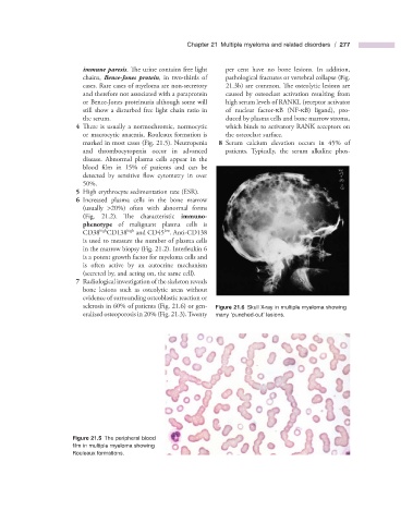
7 Radiological investigation of the skeleton reveals
bone lesions such as osteolytic areas without
evidence of surrounding osteoblastic reaction or
sclerosis in 60% of patients (Fig. 21.6 ) or gen- Figure 21.6 Skull X - ray in multiple myeloma showing
eralized osteoporosis in 20% (Fig. 21.3 ). Twenty many ‘ punched - out ’ lesions.
Figure 21.5 The peripheral blood
fi lm in multiple myeloma showing
Rouleaux formations.