
Page 293 - Essential Haematology
P. 293
(a) (b)
(c) (d)
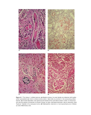
Figure 21.7 The kidney in multiple myeloma. (a) Myeloma kidney: the renal tubules are distended with hyaline
protein (precipitated light chains or Bence - Jones protein). Giant cells are prominent in the surrounding cellular
reaction. (b) Amyloid deposition: both glomeruli and several of the small blood vessels contain an amorphous
pink - staining deposit characteristic of amyloid (Congo red stain). (c) Nephrocalcinosis: calcium deposition (dark
‘ fractured ’ material) in the renal parenchyma. (d) Pyelonephritis: destruction of renal parenchyma and infi ltration
by acute infl ammatory cells.